
- Title
-
Expressed sequence tag analysis of zebrafish eye tissues for NEIBank
- Authors
- Vihtelic, T.S., Fadool, J.M., Gao, J., Thornton, K.A., Hyde, D.R., and Wistow, G.
- Source
A zebrafish eye paraffin section is overlaid with colors to highlight the tissues used for construction of the different cDNA libraries. In addition to the library made from whole eyes, libraries were also constructed using only lens or retinal tissue (shown in green or tan colors, respectively). The anterior segment library (purple) was made from tissues that included cornea (Co), annular ligament (AL), iris (Ir), the ciliary region (CR), and the retinal margin (CGZ), but did not include the lens. The posterior segment library (blue) consisted of retinal pigmented epithelium (RPE), choroid, sclera, and optic nerve (OpN) tissue, but not retina. |
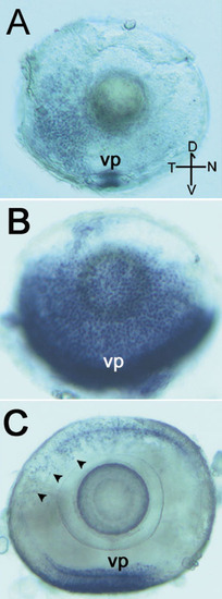
Whole-mount in situ RNA hybridizations of zebrafish cone arrestin (A,B) and rod arrestin (C) are shown. At 2.5 days post-fertilization (dpf), cone arrestin (DrArr3b) expression is highest in the ventral nasal patch (vp) of retinal cells (A). At 4 dpf, cone arrestin expression has spread into the temporal and nasal retinal regions (B). Rod arrestin (DrArr-R1) expression is identified in the ventral patch and in individual cells of the dorsal (D) retina at 4 dpf (C, arrowheads). The marker in the lower right corner of A shows the dorsal, ventral (V), nasal (N), and temporal (T) directions of the eyes. The horizontal bar of the orientation marker represents 25 μm. |

Unillustrated author statements EXPRESSION / LABELING:
|