|
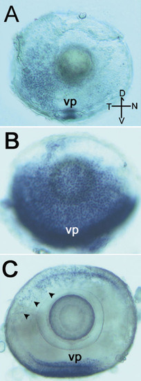
Whole-mount in situ RNA hybridizations of zebrafish cone arrestin (A,B) and rod arrestin (C) are shown. At 2.5 days post-fertilization (dpf), cone arrestin (DrArr3b) expression is highest in the ventral nasal patch (vp) of retinal cells (A). At 4 dpf, cone arrestin expression has spread into the temporal and nasal retinal regions (B). Rod arrestin (DrArr-R1) expression is identified in the ventral patch and in individual cells of the dorsal (D) retina at 4 dpf (C, arrowheads). The marker in the lower right corner of A shows the dorsal, ventral (V), nasal (N), and temporal (T) directions of the eyes. The horizontal bar of the orientation marker represents 25 μm.
|