FIGURE SUMMARY
Title

Oct4 mediates Müller glia reprogramming and cell cycle exit during retina regeneration in zebrafish

Authors
Sharma, P., Gupta, S., Chaudhary, M., Mitra, S., Chawla, B., Khursheed, M.A., Ramachandran, R.
Source
Full text @ Life Sci Alliance

ZFIN is incorporating published figure images and captions as part of an ongoing project. Figures from some publications have not yet been curated, or are not available for display because of copyright restrictions.

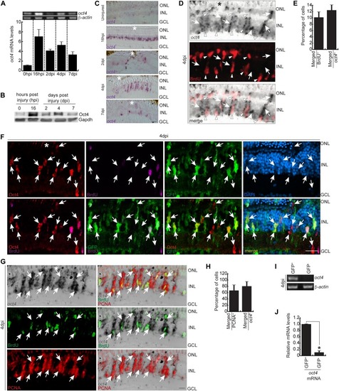
The expression pattern of Oct4, its association with MGPCs, and seclusion from BrdU<sup>+</sup> cells.

(A) RT PCR of oct4 mRNA (upper) and its qRT-PCR (lower) at various time points post retinal injury. (B) Western blot analysis of Oct4 from retinal extracts collected at different time points post injury. Gapdh is the loading control. (C) Bright-field (BF) microscopy images of retinal cross sections showing the mRNA ISH of oct4 at various time points post retinal injury. (D, E) BF and immunofluorescence (IF) confocal microscopy images of retinal cross section showing the mRNA ISH reveals the oct4 expression in the neighboring cells of BrdU+ MGPCs at 4 dpi (D), which is quantified (E). (D) White arrowheads mark BrdU+ and oct4 cells and white arrows mark oct4+ but BrdU cells in (D). (F) IF confocal microscopy images of retinal cross section, which shows the Oct4 immunofluorescence in GFP+ MGPCs in 4 dpi retina of 1016tuba1a:GFP transgenic fish. White arrows mark Oct4+ and GFP+ cells. DAPI was used as the counterstain to mark nucleus. (G, H) BF and IF confocal microscopy images of retinal cross section show the mRNA ISH of the oct4 in a significant proportion of PCNA+ MGPCs at 4 dpi (G), which is quantified (H). (G) White arrows mark PCNA+ cells that are oct4+ in (G). (I, J) RT-PCR (I) and qRT-PCR (J) of oct4 mRNA from GFP+ MGPCs compared with the GFP cells present in rest of the retina from 1016tuba1a:GFP transgenic fish at 4 dpi, *P < 0.003 (t test), N = 12. Error bars are SD. (C, D, F, G) Scale bars, 10 μm; the asterisk marks the injury site; GCL, ganglion cell layer; INL, inner nuclear layer; ONL, outer nuclear layer (C, D, F, G).

EXPRESSION / LABELING:
Genes:
Antibody:
Fish:
Condition:
Anatomical Terms:
Stage: Adult
PHENOTYPE:
Fish:
Condition:
Observed In:
Stage: Adult

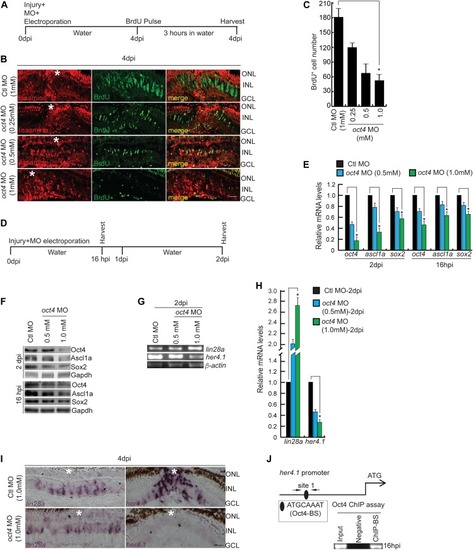
Oct4 is essential during retina regeneration.

(A) An experimental timeline that describes the MO delivery, electroporation, and BrdU pulse before harvesting at 4 dpi. (B, C) IF confocal microscopy images of retinal cross sections show the decline in BrdU+ MGPCs with increasing concentrations of oct4 MO (lissamine tag) at 4 dpi (B), which is quantified (C); *P < 0.0001 (t test), N = 4. (D) An experimental timeline that describes the MO delivery, electroporation, and harvest at 16 hpi and 2 dpi. (E) The qRT-PCR analysis of oct4, ascl1a, and sox2 genes in oct4 knockdown retina at 2 dpi and 16 hpi; *P < 0.01 (t test), N = 4. (F) Western blot analysis of Oct4, Ascl1a, and Sox2 from retinal extracts collected after oct4 knockdown at 16 hpi and 2 dpi. Gapdh is the loading control. (G, H) RT-PCR (G) and qRT-PCR (H) of lin28a and her4.1 in oct4 knockdown retina at 2 dpi. (I) BF microscopy images of retinal cross sections show the expression of lin28a and her4.1 mRNA in oct4 knockdown retina at 4 dpi. (J) The her4.1 promoter schematic reveals the typical Oct4-BS (upper) and the retinal ChIP assay confirms the physical binding of Oct4 at the typical BS (lower) in 16 hpi retina. Ctl MO is control MO. Error bars are SD. (B, I) Scale bars, 10 μm; the asterisk marks the injury site; GCL, ganglion cell layer; INL, inner nuclear layer; ONL, outer nuclear layer (B, I).

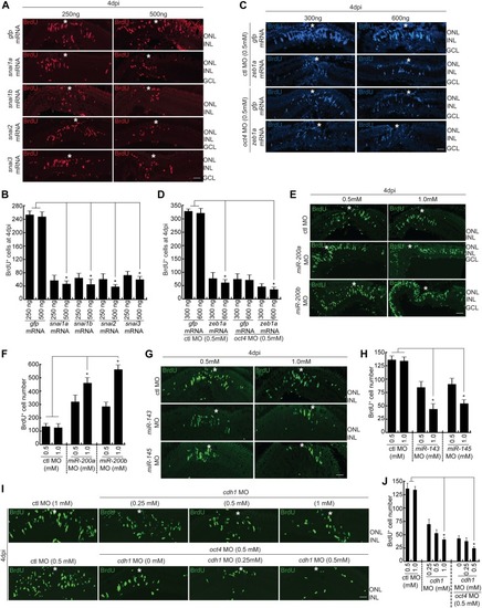
Involvement of Snails, Zebs, <italic>miR200a/miR-200b/miR-143/miR-145</italic>, and Cdh1 to ensure adequate number of MGPCs during retina regeneration.

(A, B) IF confocal microscopy images of 4 dpi retinal cross sections show BrdU+ MGPCs in snai1a, snai1b, snai2, and snai3 mRNA-transfected conditions along with gfp mRNA-transfected control retina (A), which are quantified (B); *P < 0.002 (t test), N = 4. (C, D) IF confocal microscopy images of 4 dpi retinal cross sections show BrdU+ MGPCs in zeb1a mRNA transfected conditions along with gfp mRNA transfected control retina, with control MO and oct4 MO-electroporated conditions (C), which are quantified (D); *P < 0.001 (t test), N = 4. (E, F) IF confocal microscopy images of 4 dpi retinal cross sections show BrdU+ MGPCs in miR-200a/miR-200b MO-electroporated conditions along with control MO (E), which are quantified (F); *P < 0.004 (t test), N = 4. (G, H) IF confocal microscopy images of 4 dpi retinal cross sections show BrdU+ MGPCs in miR-143/miR-145 MO-electroporated conditions along with control MO (G), which are quantified (H); *P < 0.01 (t test), N = 4. (I, J) IF confocal microscopy images of 4 dpi retinal cross sections show BrdU+ MGPCs in cdh1 MO-electroporated conditions along with control MO and oct4 MO (I), which are quantified (J); *P < 0.003 (t test), N = 4. Ctl MO is control MO. Error bars are SD. (A, C, E, G, I) Scale bars, 10 μm; the asterisk marks the injury site; GCL, ganglion cell layer; INL, inner nuclear layer; ONL, outer nuclear layer (A, C, E, G, I).

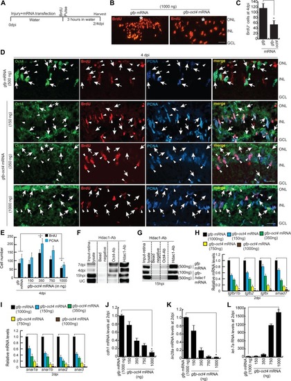
Effect of Oct4 overexpression in the injured retina.

(A) An experimental timeline that describes the mRNA transfection and BrdU pulse (for 4 dpi collection) before harvesting either at 2 or 4 dpi. (B, C) IF confocal microscopy images of retinal cross sections show reduced BrdU+ MGPCs at 4 dpi in oct4 mRNA transfected condition, compared with gfp mRNA-transfected control retina (B), which is quantified (C); *P < 0.0001 (t test), N = 4. (D) IF confocal microscopy images of retinal cross sections of oct4 mRNA-transfected retina at 4 dpi shows the cells with strong expression of Oct4 having a significant seclusion from PCNA+/BrdU+ MGPCs. White arrowheads mark BrdU+/PCNA+ cells and white arrows mark Oct4+ cells. (E) Quantification of BrdU+ and PCNA+ cells from oct4-overexpressed retina. (F) Western blot analysis of Co-IP of Oct4 and Hdac1 in retinal extracts at various time points postinjury probed with anti-Hdac1 antibody. (G) Western blot analysis of Co-IP of Oct4 and Hdac1 in retinal extracts obtained after hdac1 overexpression at 15 hpi and probed with anti-Hdac1 antibody. (H, I, J, K, L) The qRT-PCR analysis reveals the levels of tgfbr1b, tgfb2, tgfbi, smad7 (H), snails (I), cdh1 (J), lin28a (K), and let-7a miRNA (L) in oct4 mRNA-transfected retina at 2 dpi; *P < 0.001 (t test), N = 4. Error bars are SD. (B, D) Scale bars, 10 μm; the asterisk marks the injury site; GCL, ganglion cell layer; INL, inner nuclear layer; ONL, outer nuclear layer (B, D).

The increased MGPCs seen in late <italic>oct4</italic> knockdown in regenerating retina is caused by the delay in cell cycle exit.

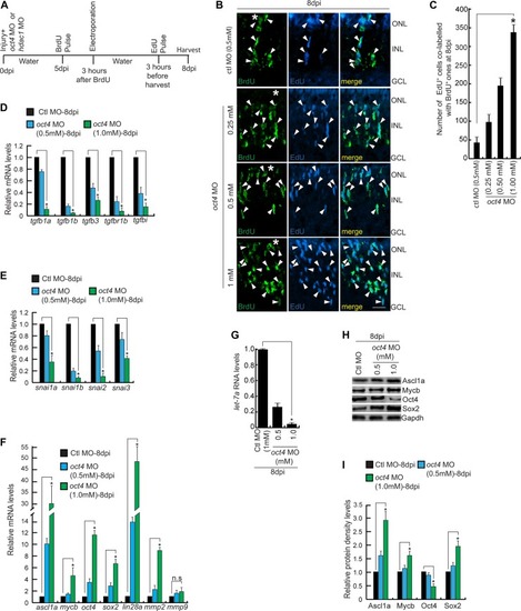
(A) An experimental timeline that describes the injury, MO injection, BrdU pulse, late electroporation of the retina, and EdU pulse 3 h before harvest at 8 dpi. (B, C) IF confocal microscopy images of retinal cross sections show increased BrdU+ MGPCs at 8 dpi in oct4 knockdown from fifth day onwards and a proof of the delay in quitting cell cycle revealed by EdU co-labeling with BrdU+ MGPCs (B), which is quantified (C); *P < 0.001, N = 4. White arrowheads mark BrdU+/EdU+ cells in (B). (D) qRT-PCR analysis of Tgf-β signaling component genes and its reporter tgfbi mRNA levels in late oct4 knockdown retina, at 8 dpi; *P < 0.02 (t test), N = 4. (E) qRT-PCR analysis of snail family genes’ mRNAs in late oct4 knockdown retina, at 8 dpi; *P < 0.03 (t test), N = 4. (F) qRT-PCR analysis of ascl1a, mycb, oct4, sox2, lin28a, mmp2, and mmp9 mRNA levels in late oct4 knockdown retina, at 8 dpi. (G) qRT-PCR analysis of let-7a miRNA levels show a decline because of oct4 late knockdown in 8 dpi retina. (H, I) Western blot analysis of various regeneration-associated factors in late oct4 knockdown retina at 8 dpi, which is quantified by densitometry (I). Gapdh is used as the loading control. Ctl MO is control MO. Error bars are SD. (B) Scale bars, 10 μm; the asterisk marks the injury site; GCL, ganglion cell layer; INL, inner nuclear layer; ONL, outer nuclear layer (B).

Effect of late <italic>hdac1</italic> and <italic>oct4</italic> knockdowns on MGPCs and gene expressions.

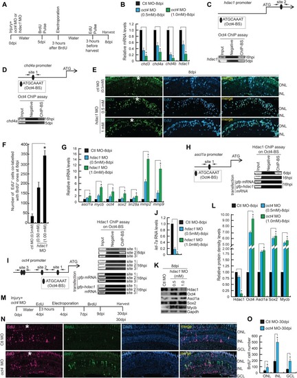
(A) An experimental timeline that describes the injury, MO injection, BrdU pulse, late electroporation of the retina, and EdU pulse 3 h before harvest at 8 dpi. (B) qRT-PCR analysis of NuRD complex component genes’ mRNA levels in late oct4 knockdown retina, at 8 dpi. (C, D) The hdac1 (C) and chd4a (D) promoter schematics reveal the typical Oct4-BSs (upper) and the retinal ChIP assays confirm the physical binding of Oct4 at these sites (lower), in 16 hpi and 5 dpi retina. (E, F) IF confocal microscopy images of retinal cross sections show increased BrdU+ MGPCs at 8 dpi in hdac1 knockdown from fifth day onwards and the delay in exiting cell cycle as revealed by EdU co-labeling with BrdU+ MGPCs (E), which is quantified (F). (G) qRT-PCR analysis of ascl1a, mycb, oct4, sox2, lin28a, mmp2, and mmp9 mRNA levels in late hdac1 knockdown retina, at 8 dpi. (H, I) The ascl1a (H) and oct4 (I) promoter schematics reveal the typical Oct4-BS (upper) and the retinal ChIP assays confirm the physical binding of Hdac1 at the Oct4-BS (lower) in 6 dpi retina. The ChIP assay performed in 16 hpi retina and also in hdac1-overexpressed condition reveal no binding of Hdac1 at Oct4-BS of ascl1a (H, right) and oct4 (I, right) promoters. The gfp mRNA transfection is the control. (J) The qRT-PCR analysis shows decreased let-7a miRNA levels with late hdac1 knockdown at 8 dpi. (K, L) Western blot analysis of different regeneration-associated factors in late hdac1 knockdown retina at 8 dpi, which is quantified by densitometry (L). Gapdh is used as the loading control. (M) An experimental timeline that describes the injury, MO injection, EdU pulse, and late electroporation of the retina at 4 dpi and BrdU on 7–9 dpi before harvest at 30 dpi. (N, O) IF confocal microscopy images of retinal cross sections show EdU and BrdU-labeled MGPCs in oct4 knockdown from the fourth day onwards and the localization of the BrdU-labeled MGPCs to various retinal layers at 30 dpi (N), which is quantified (O). Ctl MO is control MO. Error bars are SD. (E, N) Scale bars, 10 μm; the asterisk marks the injury site; GCL, ganglion cell layer; INL, inner nuclear layer; ONL, outer nuclear layer (E, N).

Increased co-localization of <italic>oct4</italic> with MGPCs towards later stages of retina regeneration, and the effect of <italic>ascl1a</italic> knockdown on <italic>oct4</italic> expression.

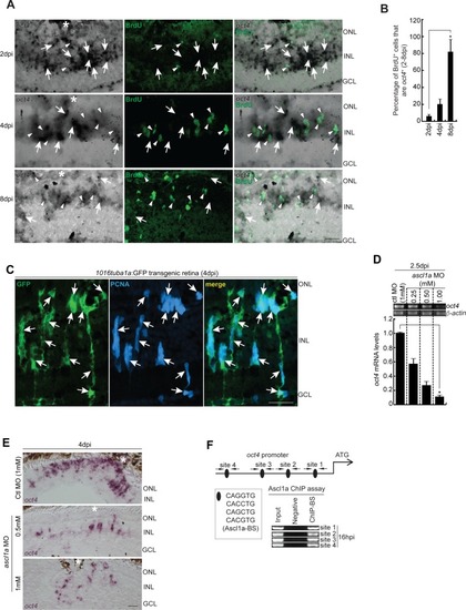
(A, B) BF and IF confocal microscopy images of retinal cross sections show increased oct4 mRNA in BrdU+ MGPCs towards the later stages of retina regeneration (A), which is quantified (B); *P < 0.0001 (t test), N = 4. (A) White arrowheads mark BrdU+ cells and white arrows mark oct4+ cells in (A). (C) IF confocal microscopy images of retinal cross sections from 1016tuba1a:GFP transgenic retina show the GFP expression in PCNA+ MGPCs at 4 dpi. White arrows mark co-labeled PCNA and GFP expression. (D) RT-PCR (upper) and qPCR (lower) analysis of oct4 mRNA in ascl1a knockdown retina at 2.5 dpi. (E) BF microscopy images of retinal cross sections show the decline in oct4 mRNA with increasing concentrations of ascl1a MO at 4 dpi. (F) The oct4 promoter schematic reveals the typical Ascl1a-BSs (upper) and the retinal ChIP assays confirm the physical binding of Ascl1a at the typical BSs (lower) in 16 hpi retina. Ctl MO is control MO. Error bars are SD. (A, C, E) Scale bars, 10 μm; the asterisk marks the injury site, GCL, ganglion cell layer; INL, inner nuclear layer; ONL, outer nuclear layer (A, C, E).

EXPRESSION / LABELING:
Gene:
Fish:
Condition:
Knockdown Reagent:
Anatomical Term:
Stage: Adult
PHENOTYPE:
Fish:
Condition:
Knockdown Reagent:
Observed In:
Stage: Adult

The reduction of MGPC proliferation in <italic>oct4</italic> knockdown retina and the rescue with <italic>gfp-oct4</italic> mRNA transfection.

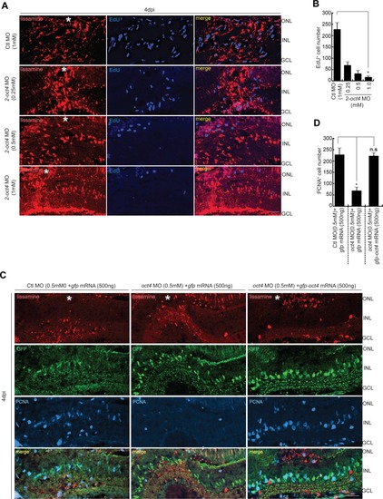
(A, B) IF confocal microscopy images of retinal cross sections show the decline in EdU+ MGPCs with increasing concentrations of 2-oct4 MO at 4 dpi (A), which is quantified (B); *P < 0.001 (t test), N = 4. (C, D) IF confocal microscopy images of retinal cross sections show the decline in PCNA+ MGPCs with oct4 MO that is rescued with gfp-oct4 mRNA at 4 dpi (C), which is quantified (D); *P < 0.001 (t test), N = 3. Ctl MO is control MO. Error bars are SD. (A, C) Scale bars, 10 μm; the asterisk marks the injury site, GCL, ganglion cell layer; INL, inner nuclear layer; ONL, outer nuclear layer (A, C).

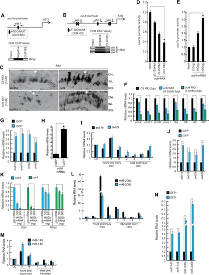
Expression dynamics of various genes reveal the Oct4 dependency during retinal regeneration.

(A, B) The promoter schematics (upper) of ascl1a (A) and oct4 (B) genes reveal the presence of Oct4-BSs, which is confirmed to be functional in a ChIP assay (lower) performed in 16 hpi retinal lysates. (C) BF confocal microscopy images of retinal cross sections show decreased ascl1a and oct4 mRNA with oct4 knockdown at 4 dpi. (D, E) The ascl1a promoter activity assayed in zebrafish embryos co-injected with ascl1a:EGFP-luciferase reporter construct and Renilla luciferase mRNA reveals a dose-dependent decrease with oct4 MO (D); *P < 0.004 (t test), N = 4 and increase with oct4 mRNA (E); *P < 0.005 (t test), N = 4, injected conditions. (F) qPCR analysis of cyclins and delta family genes in oct4 MO-electroporated retina, at 2 dpi; *P < 0.04 (t test), N = 4. (G, H) qPCR analysis of snail gene family (G) and cdh1 (H) mRNAs from GFP+ MGPCs compared with the GFP cells present in rest of the retina from 1016tuba1a:GFP transgenic fish at 4 dpi; *P < 0.004 (t test), N = 4. (I, L, M) qPCR analysis of zeb1a, zeb2a mRNA (I); miR-200a, miR-200b (L); and miR-143, miR-145 (M) miRNA levels at various time points post retinal injury. (J) qPCR analysis of zeb gene family mRNAs from GFP+ MGPCs when compared with the GFP ones present in the same retina from 1016tuba1a:GFP transgenic fish at 4 dpi; *P < 0.005 (t test), N = 4. (K) qPCR analysis of cdh1 and oct4 mRNA levels in zeb2a-transfected retina at 2 dpi and 16 hpi, compared with gfp control mRNA transfection. (N) qPCR analysis of miR-143/miR-45 and miR-200a/miR-200b miRNA levels from GFP+ MGPCs compared with the GFP cells present in rest of the retina from 1016tuba1a:GFP transgenic fish at 4 dpi; *P < 0.0001 (t test), N = 4. Ctl MO is control MO. Error bars are SD. (C) Scale bars, 10 μm; the asterisk marks the injury site, GCL, ganglion cell layer; INL, inner nuclear layer; ONL, outer nuclear layer (C).

EXPRESSION / LABELING:
Genes:
Fish:
Condition:
Knockdown Reagent:
Anatomical Term:
Stage: Adult
PHENOTYPE:
Fish:
Condition:
Knockdown Reagent:
Observed In:
Stage: Adult

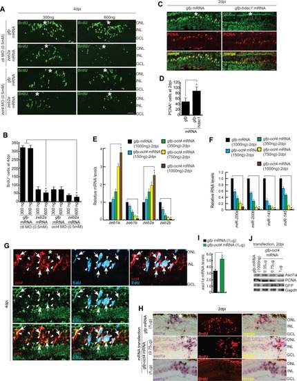
Oct4-influenced gene expression and cell proliferation during retinal regeneration.

(A, B) IF confocal microscopy images of 4 dpi retinal cross sections show BrdU+ MGPCs in zeb2a mRNA-transfected conditions along with gfp mRNA-transfected control retina, with ctl MO and oct4 MO-electroporated conditions (A), which are quantified (B); *P < 0.001 (t test), N = 4. (C, D) IF confocal microscopy images of 2 dpi retinal cross sections show PCNA+ MGPCs in hdac1 mRNA-transfected conditions along with gfp mRNA-transfected control retina (C), which is quantified (D). (E, F) qPCR analysis reveals the variations in mRNA of zeb (E), *P < 0.04 (t test), N = 4, genes, along with miR-200a, miR-200b, miR-143, and miR-145 (F), *P < 0.005 (t test), N = 4, in oct4 mRNA-transfected retina, at 2 dpi. (G) IF confocal microscopy images of retinal cross section show double-FISH of oct4 and tgfbi mRNA in 4 dpi retina. White arrowheads mark EdU+ cells and white arrows mark oct4+/tgfbi+ cells. (H, I) BF and IF microscopy images of retinal cross sections (H) and qPCR (I) show increased ascl1a and decreased BrdU with Oct4 overexpression at 2 dpi. (J) Western blot analysis of Ascl1a, PCNA, and GFP compared with Gapd from oct4 mRNA-transfected retinal extracts at 2 dpi. Ctl MO is control MO. Error bars are SD. (A, C, G, H) Scale bars, 10 μm; the asterisk marks the injury site, GCL, ganglion cell layer; INL, inner nuclear layer; ONL, outer nuclear layer (A, C, G, H).

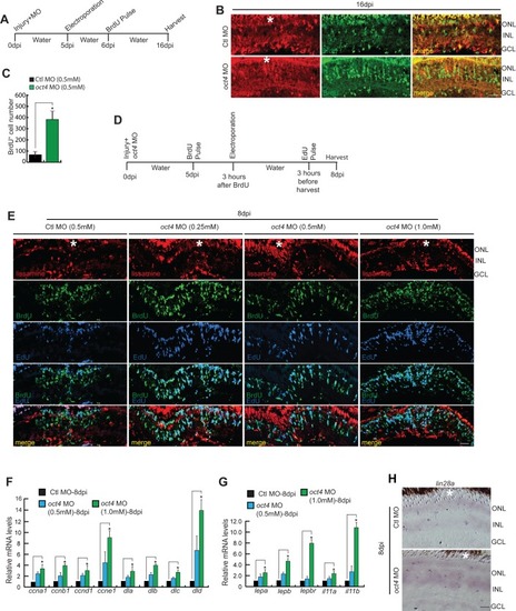
Effects of <italic>oct4</italic> late knockdown on cell proliferation and gene expression during retina regeneration.

(A) An experimental timeline that describes the injury, MO delivery, electroporation, BrdU pulse, and retina harvest at 16 dpi. (B, C) IF confocal microscopy images of retinal cross sections show increased BrdU+ MGPCs at 16 dpi in oct4 knockdown retina from fifth day onwards (B), which is quantified (C); *P < 0.007 (t test), N = 4. (D) An experimental timeline that describes the injury, MO delivery, BrdU pulse, electroporation, and retina harvest after EdU pulse at 8 dpi. (E) IF confocal microscopy images of retinal cross sections show increased BrdU+ MGPCs at 8 dpi in oct4 knockdown retina from fifth day onwards. (F, G) qPCR analysis reveals the increase in mRNA levels of cyclins and delta genes (F), *P < 0.03 (t test), N = 4, along with cytokines (G), *P < 0.04 (t test), N = 4, in late oct4 knockdown retina at 8 dpi. (H) BF microscopy images of retinal cross sections show increased lin28a levels with late oct4 knockdown at 8 dpi. Ctl MO is control MO. Error bars are SD. (B, E, H) Scale bars, 10 μm (B, E, H); the asterisk marks the injury site in (B, E, H). GCL, ganglion cell layer; INL, inner nuclear layer; ONL, outer nuclear layer (B, E, H).

EXPRESSION / LABELING:
Genes:
Fish:
Condition:
Knockdown Reagent:
Anatomical Term:
Stage: Adult
PHENOTYPE:
Fish:
Condition:
Knockdown Reagent:
Observed In:
Stage: Adult

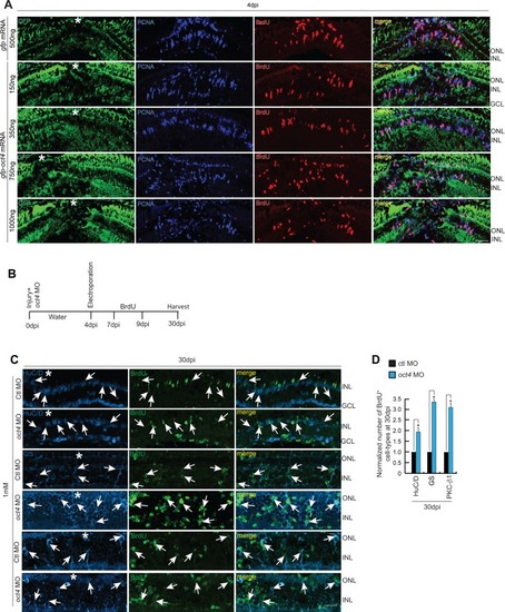
Cell proliferation in <italic>oct4</italic> overexpressed retina and cell type–specific evaluation of MGPCs with late <italic>oct4</italic> knockdown.

(A) IF confocal microscopy images of 4 dpi retinal cross sections show BrdU+ MGPCs in oct4 mRNA transfected conditions along with gfp mRNA transfected control retina. (B) An experimental timeline that describes the injury, MO delivery, electroporation, BrdU exposure, and retina harvest at 30 dpi. (C, D) IF confocal microscopy images of retinal cross sections show that the increased BrdU+ MGPCs due to late oct4 knockdown form retinal cell types at 30 dpi (C), which is quantified (D). GS, glutamine synthetase (MG cells); PKC, protein kinase C (bipolar cells); HuC/D (Amacrine cells). White arrows mark the co-labeling of different cell types with BrdU. Ctl MO is control MO. Error bars are SD. (A, C) Scale bars, 10 μm; the asterisk marks the injury site; GCL, ganglion cell layer; INL, inner nuclear layer; ONL, outer nuclear layer (A, C).

Acknowledgments
This image is the copyrighted work of the attributed author or publisher, and ZFIN has permission only to display this image to its users. Additional permissions should be obtained from the applicable author or publisher of the image. Full text @ Life Sci Alliance