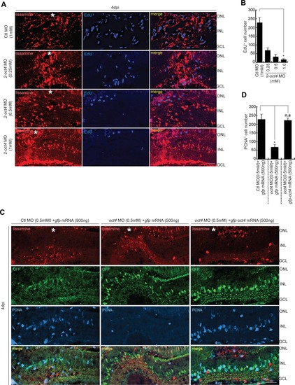
FIGURE

Figure S2.

ID
ZDB-FIG-191230-570
Publication
Sharma et al., 2019 - Oct4 mediates Müller glia reprogramming and cell cycle exit during retina regeneration in zebrafish
Other Figures
All Figure Page
Back to All Figure Page
Figure S2.

The reduction of MGPC proliferation in <italic>oct4</italic> knockdown retina and the rescue with <italic>gfp-oct4</italic> mRNA transfection.

(A, B) IF confocal microscopy images of retinal cross sections show the decline in EdU+ MGPCs with increasing concentrations of 2-oct4 MO at 4 dpi (A), which is quantified (B); *P < 0.001 (t test), N = 4. (C, D) IF confocal microscopy images of retinal cross sections show the decline in PCNA+ MGPCs with oct4 MO that is rescued with gfp-oct4 mRNA at 4 dpi (C), which is quantified (D); *P < 0.001 (t test), N = 3. Ctl MO is control MO. Error bars are SD. (A, C) Scale bars, 10 μm; the asterisk marks the injury site, GCL, ganglion cell layer; INL, inner nuclear layer; ONL, outer nuclear layer (A, C).

Expression Data

Expression Detail
Antibody Labeling
Phenotype Data

Phenotype Detail
Acknowledgments
This image is the copyrighted work of the attributed author or publisher, and ZFIN has permission only to display this image to its users. Additional permissions should be obtained from the applicable author or publisher of the image. Full text @ Life Sci Alliance