|
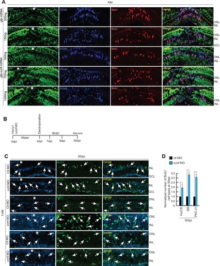
Cell proliferation in <italic>oct4</italic> overexpressed retina and cell type–specific evaluation of MGPCs with late <italic>oct4</italic> knockdown.(A) IF confocal microscopy images of 4 dpi retinal cross sections show BrdU+ MGPCs in oct4 mRNA transfected conditions along with gfp mRNA transfected control retina. (B) An experimental timeline that describes the injury, MO delivery, electroporation, BrdU exposure, and retina harvest at 30 dpi. (C, D) IF confocal microscopy images of retinal cross sections show that the increased BrdU+ MGPCs due to late oct4 knockdown form retinal cell types at 30 dpi (C), which is quantified (D). GS, glutamine synthetase (MG cells); PKC, protein kinase C (bipolar cells); HuC/D (Amacrine cells). White arrows mark the co-labeling of different cell types with BrdU. Ctl MO is control MO. Error bars are SD. (A, C) Scale bars, 10 μm; the asterisk marks the injury site; GCL, ganglion cell layer; INL, inner nuclear layer; ONL, outer nuclear layer (A, C).
|