|
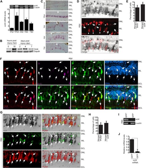
The expression pattern of Oct4, its association with MGPCs, and seclusion from BrdU<sup>+</sup> cells.(A) RT PCR of oct4 mRNA (upper) and its qRT-PCR (lower) at various time points post retinal injury. (B) Western blot analysis of Oct4 from retinal extracts collected at different time points post injury. Gapdh is the loading control. (C) Bright-field (BF) microscopy images of retinal cross sections showing the mRNA ISH of oct4 at various time points post retinal injury. (D, E) BF and immunofluorescence (IF) confocal microscopy images of retinal cross section showing the mRNA ISH reveals the oct4 expression in the neighboring cells of BrdU+ MGPCs at 4 dpi (D), which is quantified (E). (D) White arrowheads mark BrdU+ and oct4− cells and white arrows mark oct4+ but BrdU− cells in (D). (F) IF confocal microscopy images of retinal cross section, which shows the Oct4 immunofluorescence in GFP+ MGPCs in 4 dpi retina of 1016tuba1a:GFP transgenic fish. White arrows mark Oct4+ and GFP+ cells. DAPI was used as the counterstain to mark nucleus. (G, H) BF and IF confocal microscopy images of retinal cross section show the mRNA ISH of the oct4 in a significant proportion of PCNA+ MGPCs at 4 dpi (G), which is quantified (H). (G) White arrows mark PCNA+ cells that are oct4+ in (G). (I, J) RT-PCR (I) and qRT-PCR (J) of oct4 mRNA from GFP+ MGPCs compared with the GFP− cells present in rest of the retina from 1016tuba1a:GFP transgenic fish at 4 dpi, *P < 0.003 (t test), N = 12. Error bars are SD. (C, D, F, G) Scale bars, 10 μm; the asterisk marks the injury site; GCL, ganglion cell layer; INL, inner nuclear layer; ONL, outer nuclear layer (C, D, F, G).
|