
- Title
-
Interplay between Wnt2 and Wnt2bb controls multiple steps of early foregut-derived organ development
- Authors
- Poulain, M., and Ober, E.A.
- Source
- Full text @ Development
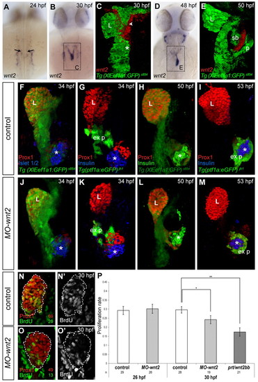
Mesodermal wnt2 controls hepatoblast proliferation. (A-E) wnt2 is expressed in bilateral LPM domains neighbouring hepatic anlage at 24 hpf (arrows, A), in the LPM abutting the proximal liver and prospective swim bladder at 30 hpf (B, arrowhead C) and confined between the swim bladder and pancreas at 50 hpf (D,E). (C,E) Projections of confocal stacks showing wnt2 mRNA (red) and Tg(XlEef1a1:GFP)s854 (green). (F-M) wnt2-depleted embryos show a mildly reduced liver at 34 hpf (J,K), which becomes more pronounced later (L,M) compared with controls (F,G,H,I). Confocal stacks showing Tg(XlEef1a1:GFP)s854 (F,H,J,L) and Tg(ptf1a:eGFP)jh1 (G,I,K,M) embryos, stained for Prox1 and Islet1/2 or Insulin, respectively. The ventral, exocrine and endocrine pancreas serve as developmental landmarks; a subset of MO-wnt2 embryos show ectopic endocrine cells (L). (N-O2) cell proliferation rates are reduced in MO-wnt2 and prt/wnt2bb embryos. Projections of confocal stacks showing 30 hpf MO-wnt2 embryos (O,O2)and controls (N,N2); numbers indicate count of individual Prox1- and BrdU-positive cells. (A-M) Dorsal views, (N-O2) ventral views; all anterior towards the top. *, endocrine pancreas; ex p, exocrine pancreas; L, liver; p, pancreas; sb, swim bladder. (P) Proliferation rates in controls, MO-wnt2 and prt/wnt2bb embryos determined by Prox1 and BrdU labelling. Standard errors are shown. Asterisks indicate the P value for each condition (*P<0.05; **P<0.001); n, number of embryos. Data are mean±s.e.m. |
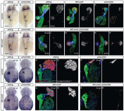
Wnt2 compensates for loss of Wnt2bb function in prt/wnt2bb mutants. (A-H) Expression of hhex at 28 hpf (A-D) and ceruloplasmin at 48 hpf (E-H) shows a reduction of liver progenitors (bracket) in embryos lacking either wnt2 (C,G) or wnt2bb (B,F) and an absence in MO-wnt2;prt/wnt2bb embryos (D,H), whereas the dorsal pancreas is unaffected (asterisk). (I-N2) Prox1 expression in Tg(XlEef1a1:GFP)s854 embryos at 52 hpf shows smaller livers in MO-wnt2 (J,J2) and prt/wnt2bb embryos (K,K2), and an increasing reduction of the liver in MO-wnt2;prt/wnt2bb embryos (L-N2), which in more severe cases includes a reduction of 2F11-positive EHPD (d:M-N2); distribution of phenotypes indicated as percentages (n=65). (O-R2) Tg(fabp10:dsRed)gz4 expression confirms the reduction of differentiating liver tissue in prt/wnt2bb embryos (P,P2) and MO-wnt2 (Q,Q2) and absence in MO-wnt2;prt/wnt2bb embryos (R,R2) at 74 hpf, indicating that the liver defect fails to recover; Tg(XlEef1a1:GFP)s854 and Tg(ela3l:EGFP)gz2) expression indicates the digestive tract and exocrine pancreas remain unaffected (O-R2). (A-H) Dorsal views and (I-R2) ventral views of projections of confocal stacks; all anterior towards the top. gb, gall bladder; L, liver; p, pancreas. EXPRESSION / LABELING:
PHENOTYPE:
|
Excess wnt2bb promotes liver formation at the expense of the ventral pancreas. (A-D2) Transient wnt2bb-overexpression in Tg(XlEef1a1:GFP)s854 (A-B2) and Tg(ptf1a:eGFP)jh1 (C-D2) embryos at 18 hpf alters foregut patterning resulting in an enlarged liver domain as visualised by Prox1 staining at 28 hpf (B, bracket: B2) and 48 hpf (D,D2). The pancreatic islet (asterisk) serves as landmark to visualize the posterior expansion of hepatoblast domain. (E-H) cp expression at 44 hpf (F) and Tg(fabp10:dsRed)gz4 expression at 74 hpf (H) indicate ectopic hepatic tissue differentiation in embryos with excess wnt2bb. Excess wnt2bb results in a severe decrease of pancreatic Tg(ptf1a:eGFP)jh1 (p: arrow, D) and Tg(ela3l:EGFP)gz2 expression (arrowhead, H). (A-D2,G,H) Ventral views of projections of confocal stacks; (E,F) dorsal views; all anterior towards the top. EXPRESSION / LABELING:
|
Ventral digestive tract endoderm posterior to the hepatic domain is competent to respond to hepatogenic signals in vivo. (A-H2) transient wnt2bb-overexpression in Tg(XlEef1a1:GFP)s854 embryos at 18 hpf leads to an expansion of the liver in posterior ventral tissues visualised by Prox1 staining at 28 hpf (A-D2) and 48 hpf (E-H2). (I,J) pdx1 expression is significantly reduced in the pancreas and anterior intestinal domains (bracket) following wnt2bb-overexpression. (L) Embryos with excess wnt2bb exhibit ectopic Prox1 and strong 2F11 staining in the EHPD domain, revealing ectopic structures (arrows), compared with controls (K). (A,C,C2,E,G,G2) dorsal views (B,D,D2,F,H,H2,K,L) ventral views of projections of confocal stacks; (I,J) dorsal views; all anterior towards the top. |
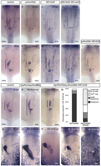
wnt2 and wnt2bb interact genetically with the receptor fzd5. (A-D) At 25 hpf, hhex-expressing liver progenitors (bracket) are absent in embryos lacking Wnt2bb (B), Fzd5 (C) or Fzd5 and Wnt2 (D), whereas the dorsal pancreas is unaffected (asterisk). (E-I) Hepatic hhex expression is reduced in prt/wnt2bb (F) and MO-fzd5 embryos (G) at 28 hpf. By contrast, in MO-wnt2;MO-fzd5 (H) or prt/wnt2bb;MO-fzd5 (I) embryos hepatoblasts fail to form. In a subset of MO-wnt2;MO-fzd5 embryos, pancreatic hhex expression is affected. (J-R) Embryos with excess wnt2bb show a significant posterior expansion of hepatic hhex or cp expression (K,O) compared with controls (J,N). In the absence of Fzd5, the majority of the embryos with excess wnt2bb exhibit no expansion of hepatic tissue (L,Q,R), whereas a subset of these embryos show a weak liver expansion (P). In the most severe cases, only single hepatoblasts form (arrow, R). (M) The distribution of liver phenotypes revealed by cp expression upon wnt2bb overexpression in wild-type or in MO-fzd5 embryos. (A-R) Dorsal views; anterior towards the top. The proportion of representative phenotype is indicated as a percentage. |
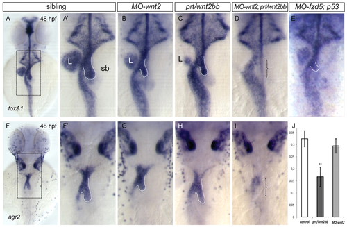
Swim bladder formation requires Wnt2, Wnt2bb and Fzd5 activity. foxa1 (A-E) and agr2 (F-I) expression at 48 hpf shows that the swim bladder (outlined by dashed line) is variably reduced in MO-wnt2 (B,G), prt/wnt2bb (C,H) MO-fzd5 (E) and absent in MO-wnt2;prt/wnt2bb embryos (bracket, D,I) compared with controls (A,A2,F,F2). Dorsal views, anterior to top. (J) Proliferation rates in controls, prt/wnt2bb and MO-wnt2 embryos determined by Tg(XlEef1a1:GFP)s854 and BrdU labelling at 42 hpf. Data are mean±s.e.m. P values are shown (**P<0.005); n>13 embryos. L, liver; sb, swim bladder. |
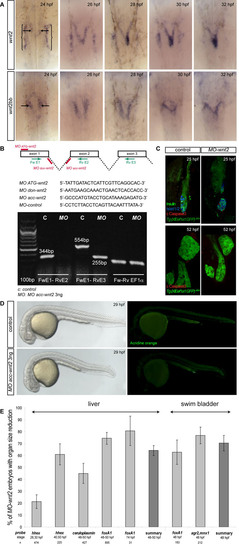
Validation of MO-wnt2. (A) wnt2 and wnt2bb mRNA expression between 24 and 32 hpf. Brackets indicate the prospective pronephros and arrows indicate wnt2 and wnt2bb expression in the LPM. wnt2 domains differ from wnt2bb expression in that they are located in more medial and posterior regions of the mesoderm. (B) The wnt2 knock-down strategy; sequences of the relevant MOs are shown. RT-PCR analysis MO acc-wnt2 embryos show a complete knock-down of Wnt2, as indicated by a loss of exon2; sequences of indicated PCR-primers are: Fw E1, 52-CGTAGACAAGTGCCTGAACG-32 Rv E2, 52-CAGGAGCCTCCCGAACAG-32 and Rv E3, 52-GACCTGGGTGAACTTGATGG-32. (C) MO-wnt-2 embryos display no apparent cell death in the foregut endoderm at 25 and 52 hpf, revealed by staining for cleaved caspase 3 in Tg(XlEef1a1:GFP)s854 embryos. Islet1/2, insulin-positive dorsal pancreas serves as landmark. (D) Likewise, Acridine Orange staining shows no overall increase of cell death at 29 hpf in MO-wnt2 embryos. (E) Quantification of whole-mount mRNA expression analyses reveals 64% of MO-wnt2 embryos show a hypoplastic liver and 70% a hypoplastic swim bladder; standard errors are indicated; n, number of embryos. (A) Dorsal views, (C) 27 hpf and 52 hpf, single confocal section of ventral view; all anterior towards the top. (D) Lateral views, anterior towards the left. |
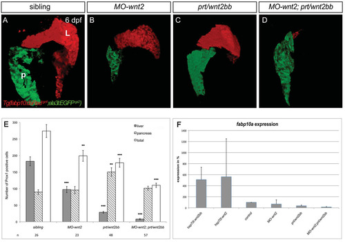
wnt2 and wnt2bb are essential for liver formation. (A-D) Projections of confocal stacks showing ventral views of Tg(fabp10:dsRedgz4;ela3l:EGFPgz2) embryos, anterior to the top; L, liver; P, pancreas. MO-wnt2 (B) or prt/wnt2bb (C) livers have partially recovered in size at 6 dpf, whereas 23% of MO-wnt2;prt/wnt2bb embryos fail to differentiate hepatic tissue and 46% of embryos show only few hepatic cells (D; n=13). (E) The number of liver and ventral pancreas cells determined by counting Prox1-positive nuclei at 48 hpf. Sample number, standard errors and P values are shown (*P<0.05; **P<0.005; ***P<0.0005). (F) qPCR quantification of fabp10a expression levels at 48 hpf from embryos lacking Wnt2bb, Wnt2 or both ligands, as well as from embryos in which either ligand has been transiently activated at 18 hpf. Importantly, expression levels in prt/wnt2bb and MO-wnt2;prt/wnt2bb are at the limit of detection and therefore probably negligible. Results represent an average of two independent experiments and standard deviations are shown. |
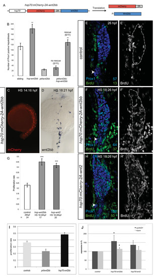
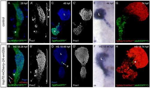
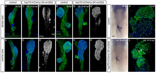
Excess wnt2 or wnt2bb promotes hepatoblast specification and proliferation. (A) The wnt2bb overexpression construct. The hsp70 promoter controls expression of a mCherry-2A-wnt2bb transcript, which upon translation produces independent mCherry and Wnt2bb protein, owing to an unstable bond in the viral 2A-linker peptide. (B) The phenotypic rescue in a subset of prt/wnt2bb embryos transiently overexpressing wnt2bb, determined by hepatic Prox1 expression. (C) Live picture of injected embryo 2 hours after heat-shock. mCherry indicates cells in which the hsp70:mCherry-2A-wnt2bb construct was activated. (D) Whole-mount mRNA in situ hybridization for wnt2bb showing ectopic activation 3 hours after heat-shock. Staining was stopped before endogenous expression was detectable. (E-H2) Cell proliferation rates are increased in embryos in which wnt2bb (F,F2) or wnt2 (H,H2) have been ectopically activated; numbers indicate count of individual Prox1- and BrdU-positive cells. Liver anlage (broken line) is expanded posteriorly in embryos with excess wnt2bb (F,F2) and wnt2 (H,H2) compared with controls (E,E2); inter-renal primordium (arrowhead) serves as landmark. (G) Proliferation rates in controls, hsp70:mCherry-2A-wnt2bb and hsp70:mCherry-2A-wnt2 embryos determined by Prox1 and BrdU labelling. (I) Cell proliferation in the LPM is decreased in prt/wnt2bb embryos at 30 hpf and increased in embryos after its transient activation at 18 hpf; proliferating cells were determined by BrdU and Tg(XlEef1a1:GFP)s854 to exclude the endoderm. (J) cyclinD1 and myca expression 4 hours after transient wnt2bb and wnt2 overexpression at 26 hpf; controls represent siblings exposed to the same heat-shock regime to determine the effect specific to Wnt overexpression. (C) Lateral view, (D) dorsal view, (E-H2) ventral views of projections of confocal stacks; all anterior towards the top. (B,G,I,J) Standard errors and P values are shown (*Pd0.05; **P<0.001; ***P<2.10-8). |
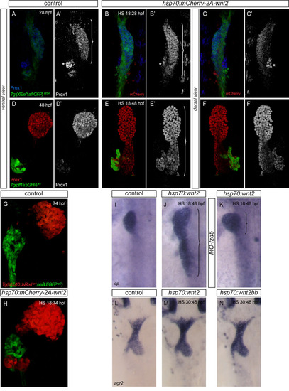
Excess wnt2 expression promotes liver formation at the expense of pancreatic tissue. (A-F2) transient wnt2-overexpression in Tg(XlEef1a1:GFP)s854 (A-C2) and Tg(ptf1a:eGFP)jh1 (D-F2) at 18 hpf alters foregut patterning resulting in an enlarged liver domain as visualised by Prox1 staining at 28 hpf (B, bracket in B2) and 48 hpf (E,E2). The inter-renal primordium (asterisk) serves as landmark to visualize the posterior expansion of the hepatoblast domain. This expansion is confined to the ventral digestive tract endoderm (C,C2,F,F2). (G,H) Tg(fabp10:dsRed)gz4 expression at 74 hpf indicates ectopic hepatic tissue differentiation in embryos with excess wnt2 (H). Excess wnt2 results in a significant decrease of pancreatic Tg(ptf1a:eGFP)jh1 (E,F) and Tg(ela3l:eGFP)gz2 (H) expression. (I-K) In embryos lacking Fzd5, the majority of embryos display no posterior expansion of hepatic cp expression at 48 hpf after transient wnt2 expression at 18 hpf (K). (L-N) agr2 expression at 48 hpf indicates no apparent alteration of swim bladder formation upon transient overexpression of wnt2 or wnt2bb at 30 hpf (M and N, respectively). (A-B2,D-E2,G,H) Ventral views of projections of confocal stacks, (C,C2,F,F2,I-N) dorsal views; all anterior towards the top. |
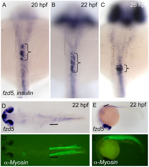
Endodermal frizzled5 expression includes the presumptive hepatic domain at the time of liver specification. (A-E2) fzd5 is widely expressed in the endoderm at 20 (A), 22 (B,D-E2) and 26 (C) hpf, including the presumptive hepatic domain (dashed bracket) anterior to the endocrine islet and the more posteriorly located intestine. Insulin-expressing endocrine islet cells (dark, high-level expressing cells; plain bracket) serve as anteroposterior landmark. (D-E2) endodermal fzd5 expression includes the hepatic endoderm, located at the height of somites 1-3 (black line), which are visualized by α-Myosin staining (D2,E2). (A-D2) Dorsal views, (E,E2) lateral view; (A-C) anterior is towards the top; (D-E2) anterior is towards the left. |