
|
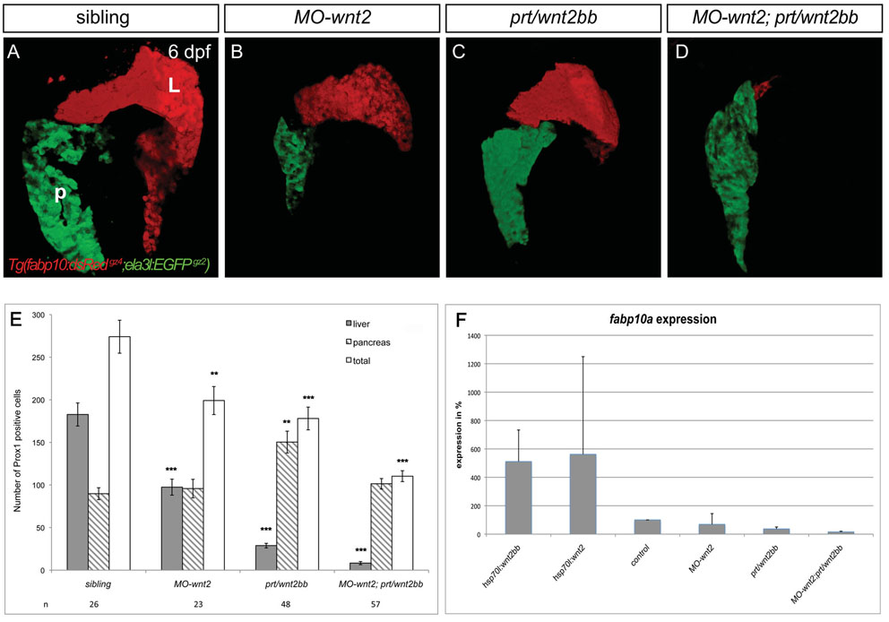
Fig. S2
wnt2 and wnt2bb are essential for liver formation. (A-D) Projections of confocal stacks showing ventral views of Tg(fabp10:dsRedgz4;ela3l:EGFPgz2) embryos, anterior to the top; L, liver; P, pancreas. MO-wnt2 (B) or prt/wnt2bb (C) livers have partially recovered in size at 6 dpf, whereas 23% of MO-wnt2;prt/wnt2bb embryos fail to differentiate hepatic tissue and 46% of embryos show only few hepatic cells (D; n=13). (E) The number of liver and ventral pancreas cells determined by counting Prox1-positive nuclei at 48 hpf. Sample number, standard errors and P values are shown (*P<0.05; **P<0.005; ***P<0.0005). (F) qPCR quantification of fabp10a expression levels at 48 hpf from embryos lacking Wnt2bb, Wnt2 or both ligands, as well as from embryos in which either ligand has been transiently activated at 18 hpf. Importantly, expression levels in prt/wnt2bb and MO-wnt2;prt/wnt2bb are at the limit of detection and therefore probably negligible. Results represent an average of two independent experiments and standard deviations are shown.