
|
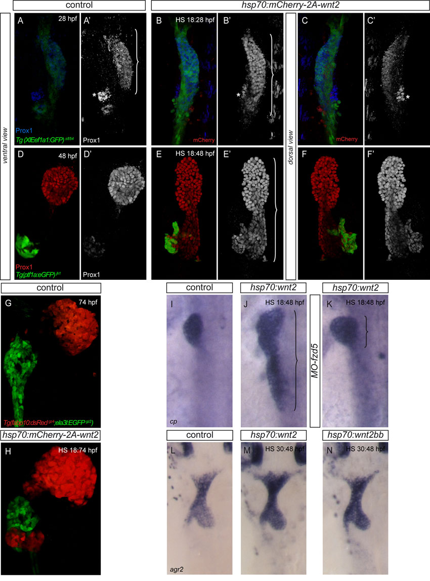
Fig. S4
Excess wnt2 expression promotes liver formation at the expense of pancreatic tissue. (A-F2) transient wnt2-overexpression in Tg(XlEef1a1:GFP)s854 (A-C2) and Tg(ptf1a:eGFP)jh1 (D-F2) at 18 hpf alters foregut patterning resulting in an enlarged liver domain as visualised by Prox1 staining at 28 hpf (B, bracket in B2) and 48 hpf (E,E2). The inter-renal primordium (asterisk) serves as landmark to visualize the posterior expansion of the hepatoblast domain. This expansion is confined to the ventral digestive tract endoderm (C,C2,F,F2). (G,H) Tg(fabp10:dsRed)gz4 expression at 74 hpf indicates ectopic hepatic tissue differentiation in embryos with excess wnt2 (H). Excess wnt2 results in a significant decrease of pancreatic Tg(ptf1a:eGFP)jh1 (E,F) and Tg(ela3l:eGFP)gz2 (H) expression. (I-K) In embryos lacking Fzd5, the majority of embryos display no posterior expansion of hepatic cp expression at 48 hpf after transient wnt2 expression at 18 hpf (K). (L-N) agr2 expression at 48 hpf indicates no apparent alteration of swim bladder formation upon transient overexpression of wnt2 or wnt2bb at 30 hpf (M and N, respectively). (A-B2,D-E2,G,H) Ventral views of projections of confocal stacks, (C,C2,F,F2,I-N) dorsal views; all anterior towards the top.