
Fig. S4
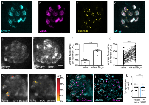
SypHy localizes to synaptic vesicles, and it is properly acidified; active and silent cells have the same number of synapses. (a-d) Immunostaining of a representative neuromast expressing SypHy. Merged image (d) shows SypHy (a, cyan) colocalizes with Vglut3 (b, magenta) near Ribeye b labeled presynaptic ribbons (c, yellow). (e) Example neuromast demonstrating that baseline SypHy signals (e, left panel) increase when SypHy is deacidified after 40 mM NH4 Cl treatment (e, right panel). (f-g) Quantification of baseline SypHy intensities after 40 mM NH4 Cl treatment. The average SypHy signal per neuromast (f) is increased (naïve: 298.00 a.u. ± 6.87; after 40 mM NH4 +: 388.40 a.u. ± 10.62, n = 6 neuromasts, p = 0.0007). All hair cells show an increase in baseline SypHy signal (g), (naïve: 297.40 a.u. ± 5.42; after 40 mM NH4 +: 387.30 a.u. ± 6.53, n = 85 cells, p < 0.0001). (h) SypHy signals in response to 2-s anterior (h, left panel) and 2-s posterior (h, right panel) step stimuli that cumulatively activate all hair cells. Spatial patterns of SypHy signals during stimulation are colorized according to the ΔF/F heat maps and superimposed onto a pre-stimulus baseline image. (i) Live image of hair cells from the same neuromast as (h) expressing SypHy (cyan) and Ribeye b-mCherry (magenta). (j) The same neuromast as in (h-i), after immunostaining to label postsynaptic MAGUK (cyan) and presynaptic Ribeye b (magenta). White circles mark hair cells with vesicle fusion and blue circles indicate cells with no vesicle fusion. (k) Quantification of the number of synapses per individual hair cell based on MAGUK and Ribeye b staining show similar number of synapses between cells with (3.20 ± 1.18, n = 15 cells) and without (3.05 ± 0.20, n = 22 cells) vesicle fusion, p = 0.39, day 6 4-5. A paired t-test was used in (f-g), and a Mann-Whitney test was used in (k), ***p < 0.001, ****p < 0.0001. Scale bars = 5 μm. |