
Fig. S1
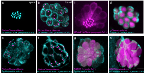
Zebrafish transgenic lines for functional imaging. (a-b) Top-down view of a double transgenic neuromast expressing a membrane localized Ca2+ indicator, GCaMP6s-CAAX (cyan) and Ribeye b-mCherry (magenta) to label presynaptic ribbons in hair cells. An apical GCaMP6s-CAAX plane is used to examine mechanosensative Ca2+ influx in the mechanosensory hair bundles (a) while a plane at the base of the same neuromast can be used to monitor presynaptic Ca2+ influx at ribbons (b). (c) Similar to GCaMP6s-CAAX, red-shifted jRCaMP1a-CAAX (magenta) can measure apical or basal Ca2+ influx in hair cells. (d) Bongworri (cyan) can be used to detect membrane voltage changes in hair cells. (e) A double transgenic line expressing SypHy (cyan), an indicator of vesicle fusion and Ribeye b-mCherry (magenta) in hair cells can be used to monitor vesicle fusion at presynaptic ribbons. (f) Postsynaptic afferents expressing GCaMP6s-CAAX (cyan), along with expression of Ribeye b-mCherry (magenta) in hair cells enables monitoring of postsynaptic Ca2+ signals adjacent to presynaptic ribbons. (g) Cells expressing SypHy (cyan) and RGECO1 (magenta), a red cytosolic Ca2+ indicator enable dual monitoring of hair-cell Ca2+ and vesicle fusion. (h) A double transgenic with postsynaptic afferents expressing GCaMP6s-CAAX (cyan) and hair cells expressing RGECO1 (magenta) enable monitoring of pre- and post-synaptic Ca2+ activities respectively. Scale bar = 5 μm. |