
- Title
-
Cardiac myocyte diversity and a fibroblast network in the junctional region of the zebrafish heart revealed by transmission and serial block-face scanning electron microscopy
- Authors
- Lafontant, P.J., Behzad, A.R., Brown, E., Landry, P., Hu, N., and Burns, A.R.
- Source
- Full text @ PLoS One
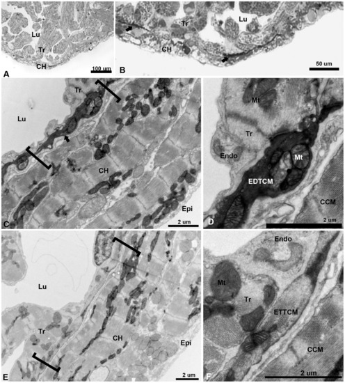
Low magnification of the adult zebrafish heart by light and TEM. (A), light microscopy view of the apical region of the zebrafish ventricle showing a thin layer compact heart (CH) with trabeculae (Tr) projecting within the lumen (Lu). (B), higher magnification of the apex showing darkly stained linear structures (arrows) close to the spongy-compact interface of the ventricular myocardium. (C), low magnification TEM of compact ventricular myocardium constituted of four to five overlapping cardiac myocytes (CM) layers from epicardium (Epi) to lumen (Lu), with electron dense cells (arrow) within a complex junctional region (JR, brackets) at the interface of compact and spongy myocardium. (D), higher magnification show mitochondria (Mt) filled section of electron dense transitional cardiac myocyte (EDTCM). EDTCM are apposed to trabecular cardiac myocytes and endocardial cells (Endo) on the luminal side. EDTCMs are separated from the compact cardiac myocytes (CCM) by a junctional space, an interstitial space within the JR. (E), low magnification TEM of the JR (brackets) showing flattened electron translucent transitional cardiac myocytes (ETTCM) in contact with trabeculae and in proximity to endocardium (Endo) on the luminal side. (F), higher magnification of a flattened cells show presence of actin and myosin filaments, apposition to trabeculae and endocardial cells as well as separation from the compact myocardium by the junctional space. |
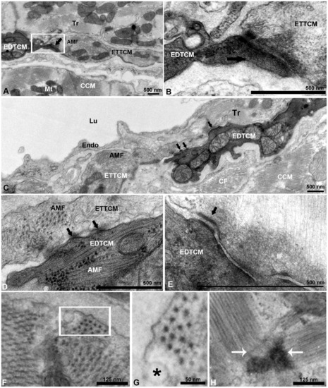
Electron dense and electron translucent transitional cardiac myocytes interactions. (A), example of end-to-end interaction between EDTCM and electron translucent cardiac myocytes (ETTCM) forming a continuum at the spongy-compact interface. (B), higher magnification of contact area with complex adhesion junctions including desmosomes between the two cells. (C), example of interaction between mitochondria filled and actin-myosin filament (AMF) containing EDTCM cells and ETTCM via adhesion junctions (arrows). (D), higher magnification of EDTCM and ETTCM showing gap junctions (arrows), and (E), desmosomes. (F), AMF in a transitional cell. (G), higher magnification (inset in F, rotated) showing the hexagonal array of myosin in cardiac myocytes. (F) sarcomere and z-band in the same cell. AMF, actin-myosin filament; CCM, compact cardiac myocytes, CM, cardiac myocytes; EDTCM, electron dense transitional cardiac myocytes; ETTCM, electron translucent transitional cardiac myocytes; Endo, endocardium; Epi; IF, intermediate filaments; Lu, lumen; Mt, mitochondrion; Tr, trabeculae. |
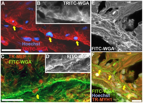
Immunofluorescence imaging of the zebrafish ventricle junctional region. (A), staining with TRITC-labeled wheat germ agglutinin (WGA) highlighting cardiac myocytes borders, in conjunction with Hoechst staining, to illustrate the transitional cardiac myocytes (arrows). (B), original monochrome image of the TRITC-WGA stained cell borders containing the two right-most nuclei identified by the arrows in panel A. (C), immunostaining with MEF antibody to label myocyte nuclei and FITC-labeled WGA-stained cell membranes; the arrow identifies a transitional cardiac myocyte. (D), original monochrome image of the FITC-WGA stained cell membranes in panel C around the nucleus identified by the arrow. (E), confocal image of FITC-WGA-stained cardiac myocyte membranes. (F), overlay images of FITC-WGA in panel E with MYH1 (anti-myosin heavy chain-1 antibody) immunostaining, and Hoechst staining identifying transitional cardiac myocytes (arrows). Scale bar, 10 μm. |
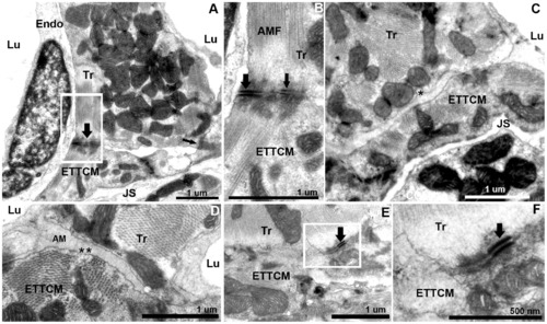
Transitional cardiac myocytes ring and trabecular cardiac myocytes contacts. (A), trabecular cardiac myocytes (Tr) in direct contact with two CM of the transitional ring. Trabeculae and transitional CMs are quasi-perpendicularly oriented. Contacts are mediated by electron dense adhesion junctions (arrows). (B), higher magnification of (A), with fascia adherentes junction (thick arrow) and desmosome (thin arrow) between the Tr and ETTCM. Well organized actin and myosin filaments are oriented at an approximately 145 degree angle. (C, D), region of approximation of trabecular CM and transitional CM with amorphous material (AM) interposed between the two cell membranes. Note a number of caveolae in the trabecular CM membrane (C,*) on their abluminal side, and in the transitional CM membrane (D,*) on the luminal side. (E), another example of desmosome linking a trabecular to a transitional CM. (F), higher magnification of (E). |
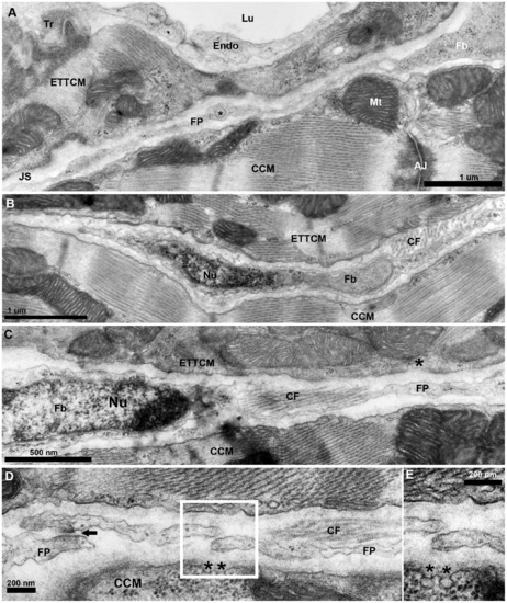
Fibroblasts network in the junctional region of the zebrafish ventricle. (A), TEM of a fibroblast (Fb) cytoplasm and its thin extended process/filopodium (FP) 100 to 200 nm thick spanning the junctional space (JS) in between the transitional CM ring and the adjacent compact cardiac myocyte (CCM). Note vesicle (*) in an enlarged region (~300 nm) of the fibroblasts filopodium (FP). (B), fibroblast profile with nucleus (Nu) and its cytoplasm extending into a long thin process, and collagen fiber (CF) bundles running perpendicular to the section’s plane. (C), another fibroblast and its nucleus (Nu) and with cytoplasm extending into a long process, and collagen fiber (CF) bundles running parallel to the section’s plane. (D), fibroblast filopodial termini in close approximation. Electron dense region in the close approximation area suggestive of specialized adhesion junction (arrow). Note the presence of caveolae (*) on the membrane of the CCM facing the fibroblast in the junctional space. |
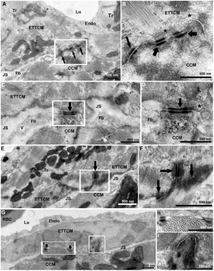
Transitional CM ring and compact CM contacts. (A), ETTCM and Compact CM direct contacts mediated by adhesion junctions (arrows). Note the JS with fibroblast (Fb) on the right and narrow JS of the left of the contact region. (B), higher magnification of inset in (A) showing adhesion junctions associated with fascia adherentes (thick arrow), and also with desmosomes (thin arrow). Note the abundance of caveolae (*) in the transitional CM membrane in the contact region. (C), another narrow region of contact (arrow) between a transitional and a compact cardiac myocyte forming a bridge between the two cells and adjacent Z-band in the CCM. A fibroblast process is seen on each side of the junction. Note a vesicle (V) in the fibroblast process. (D), higher magnification of the contact area (arrow) showing a desmosome associated with actin, myosin, and intermediate filaments on the ETTCM and abundant ribosomes (rb) in the CCM. (E), a tri-cellular junction between two transitional cardiac CM and one compact CM. (F), higher magnification of (E, inset) showing desmosomes between the ETTCM and each ETTCM with the compact CM (arrows). (G), discrete adhesion junction between a transitional and compact CM (arrows). (H), higher magnification of inset 1 of (F). (I), higher magnification of inset 2 of F, showing compact CM contact within an invagination of the transitional CM. |
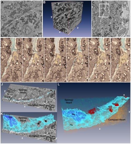
Segmentation and three-dimensional reconstruction of the junctional region of zebrafish ventricle. (A), the first 25×25 um image of 800 images acquired in a region of interest with fibroblasts running diagonally across the image plane, and a vessel profile in the lower half. (B), three-dimensional reconstruction using Amira 5.2 of 800 25×25 um sections at 50 nm intervals demonstrating the 3D spread of fibroblasts in the junctional space. The vessel in Panel A is seen projected in the yz plane in the compact myocardium. (C), a single 25×25 um image of 500 images from another region of interest with inset containing a fibroblast nucleus and fibroblasts processes within the junctional space. (D–I), six serial sections of panel C inset region, 100 nm apart, with fibroblasts (highlighted, blue) in the junctional space and a cardiac myocyte (yellow) from the compact heart. (D–F), fibroblasts can be seen occupying the uninterrupted junctional space. (G), shows the cardiac myocyte progressively protruding into the junctional space while fibroblasts processes are parted in the plane of observation. (H–I), the compact heart cardiac myocyte makes contact with the spongy heart cardiac myocyte. (J), single section in the yz plane with fibroblasts (Fb) highlighted in light blue. (K), following segmentation of 60 serial sections in Amira, 3D reconstruction of fibroblasts (light blue) demonstrates their sheet-like structure; the segmented nucleus (Nu) can be seen (dark blue). Two openings are revealed in the fibroblasts sheet (*); these openings correspond to the location of compact-transitional ring myocytes contacts. (L), Additional segmentation of the transitional cardiac myocytes in 25×25×6 um volume, showing the sheet-like appearance of fibroblasts within the junctional region. Four contact surfaces between compact and transitional cardiac myocytes are revealed (red) through openings in the fibroblasts network. |
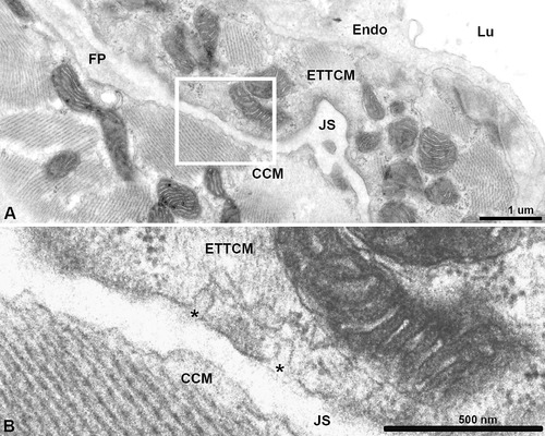
Transitional myocyte abluminal caveolae. (A), Junctional space (JS) with a fibroblast process (FP). (B), Higher magnification of inset in (A) showing caveolae (*) on transitional CM facing the JS. |