
Fig. 4
- ID
- ZDB-FIG-140115-8
- Publication
- Lafontant et al., 2013 - Cardiac myocyte diversity and a fibroblast network in the junctional region of the zebrafish heart revealed by transmission and serial block-face scanning electron microscopy
- Other Figures
- All Figure Page
- Back to All Figure Page
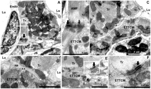
Transitional cardiac myocytes ring and trabecular cardiac myocytes contacts. (A), trabecular cardiac myocytes (Tr) in direct contact with two CM of the transitional ring. Trabeculae and transitional CMs are quasi-perpendicularly oriented. Contacts are mediated by electron dense adhesion junctions (arrows). (B), higher magnification of (A), with fascia adherentes junction (thick arrow) and desmosome (thin arrow) between the Tr and ETTCM. Well organized actin and myosin filaments are oriented at an approximately 145 degree angle. (C, D), region of approximation of trabecular CM and transitional CM with amorphous material (AM) interposed between the two cell membranes. Note a number of caveolae in the trabecular CM membrane (C,*) on their abluminal side, and in the transitional CM membrane (D,*) on the luminal side. (E), another example of desmosome linking a trabecular to a transitional CM. (F), higher magnification of (E). |