
FIGURE
Fig. S1
- ID
- ZDB-FIG-140115-12
- Publication
- Lafontant et al., 2013 - Cardiac myocyte diversity and a fibroblast network in the junctional region of the zebrafish heart revealed by transmission and serial block-face scanning electron microscopy
- Other Figures
- All Figure Page
- Back to All Figure Page
Fig. S1
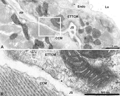
Transitional myocyte abluminal caveolae. (A), Junctional space (JS) with a fibroblast process (FP). (B), Higher magnification of inset in (A) showing caveolae (*) on transitional CM facing the JS. |
Expression Data
Expression Detail
Antibody Labeling
Phenotype Data
Phenotype Detail
Acknowledgments
This image is the copyrighted work of the attributed author or publisher, and
ZFIN has permission only to display this image to its users.
Additional permissions should be obtained from the applicable author or publisher of the image.
Full text @ PLoS One