
Fig. 1
- ID
- ZDB-FIG-140115-5
- Publication
- Lafontant et al., 2013 - Cardiac myocyte diversity and a fibroblast network in the junctional region of the zebrafish heart revealed by transmission and serial block-face scanning electron microscopy
- Other Figures
- All Figure Page
- Back to All Figure Page
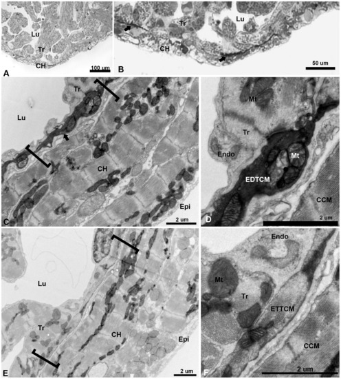
Low magnification of the adult zebrafish heart by light and TEM. (A), light microscopy view of the apical region of the zebrafish ventricle showing a thin layer compact heart (CH) with trabeculae (Tr) projecting within the lumen (Lu). (B), higher magnification of the apex showing darkly stained linear structures (arrows) close to the spongy-compact interface of the ventricular myocardium. (C), low magnification TEM of compact ventricular myocardium constituted of four to five overlapping cardiac myocytes (CM) layers from epicardium (Epi) to lumen (Lu), with electron dense cells (arrow) within a complex junctional region (JR, brackets) at the interface of compact and spongy myocardium. (D), higher magnification show mitochondria (Mt) filled section of electron dense transitional cardiac myocyte (EDTCM). EDTCM are apposed to trabecular cardiac myocytes and endocardial cells (Endo) on the luminal side. EDTCMs are separated from the compact cardiac myocytes (CCM) by a junctional space, an interstitial space within the JR. (E), low magnification TEM of the JR (brackets) showing flattened electron translucent transitional cardiac myocytes (ETTCM) in contact with trabeculae and in proximity to endocardium (Endo) on the luminal side. (F), higher magnification of a flattened cells show presence of actin and myosin filaments, apposition to trabeculae and endocardial cells as well as separation from the compact myocardium by the junctional space. |