
Fig. 5
- ID
- ZDB-FIG-140115-9
- Publication
- Lafontant et al., 2013 - Cardiac myocyte diversity and a fibroblast network in the junctional region of the zebrafish heart revealed by transmission and serial block-face scanning electron microscopy
- Other Figures
- All Figure Page
- Back to All Figure Page
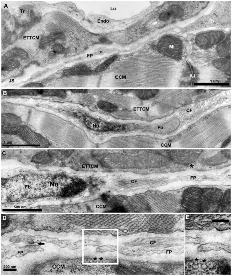
Fibroblasts network in the junctional region of the zebrafish ventricle. (A), TEM of a fibroblast (Fb) cytoplasm and its thin extended process/filopodium (FP) 100 to 200 nm thick spanning the junctional space (JS) in between the transitional CM ring and the adjacent compact cardiac myocyte (CCM). Note vesicle (*) in an enlarged region (~300 nm) of the fibroblasts filopodium (FP). (B), fibroblast profile with nucleus (Nu) and its cytoplasm extending into a long thin process, and collagen fiber (CF) bundles running perpendicular to the section’s plane. (C), another fibroblast and its nucleus (Nu) and with cytoplasm extending into a long process, and collagen fiber (CF) bundles running parallel to the section’s plane. (D), fibroblast filopodial termini in close approximation. Electron dense region in the close approximation area suggestive of specialized adhesion junction (arrow). Note the presence of caveolae (*) on the membrane of the CCM facing the fibroblast in the junctional space. |