
Fig. 7
- ID
- ZDB-FIG-140115-11
- Publication
- Lafontant et al., 2013 - Cardiac myocyte diversity and a fibroblast network in the junctional region of the zebrafish heart revealed by transmission and serial block-face scanning electron microscopy
- Other Figures
- All Figure Page
- Back to All Figure Page
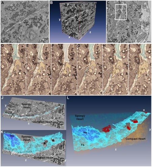
Segmentation and three-dimensional reconstruction of the junctional region of zebrafish ventricle. (A), the first 25×25 um image of 800 images acquired in a region of interest with fibroblasts running diagonally across the image plane, and a vessel profile in the lower half. (B), three-dimensional reconstruction using Amira 5.2 of 800 25×25 um sections at 50 nm intervals demonstrating the 3D spread of fibroblasts in the junctional space. The vessel in Panel A is seen projected in the yz plane in the compact myocardium. (C), a single 25×25 um image of 500 images from another region of interest with inset containing a fibroblast nucleus and fibroblasts processes within the junctional space. (D–I), six serial sections of panel C inset region, 100 nm apart, with fibroblasts (highlighted, blue) in the junctional space and a cardiac myocyte (yellow) from the compact heart. (D–F), fibroblasts can be seen occupying the uninterrupted junctional space. (G), shows the cardiac myocyte progressively protruding into the junctional space while fibroblasts processes are parted in the plane of observation. (H–I), the compact heart cardiac myocyte makes contact with the spongy heart cardiac myocyte. (J), single section in the yz plane with fibroblasts (Fb) highlighted in light blue. (K), following segmentation of 60 serial sections in Amira, 3D reconstruction of fibroblasts (light blue) demonstrates their sheet-like structure; the segmented nucleus (Nu) can be seen (dark blue). Two openings are revealed in the fibroblasts sheet (*); these openings correspond to the location of compact-transitional ring myocytes contacts. (L), Additional segmentation of the transitional cardiac myocytes in 25×25×6 um volume, showing the sheet-like appearance of fibroblasts within the junctional region. Four contact surfaces between compact and transitional cardiac myocytes are revealed (red) through openings in the fibroblasts network. |