
|
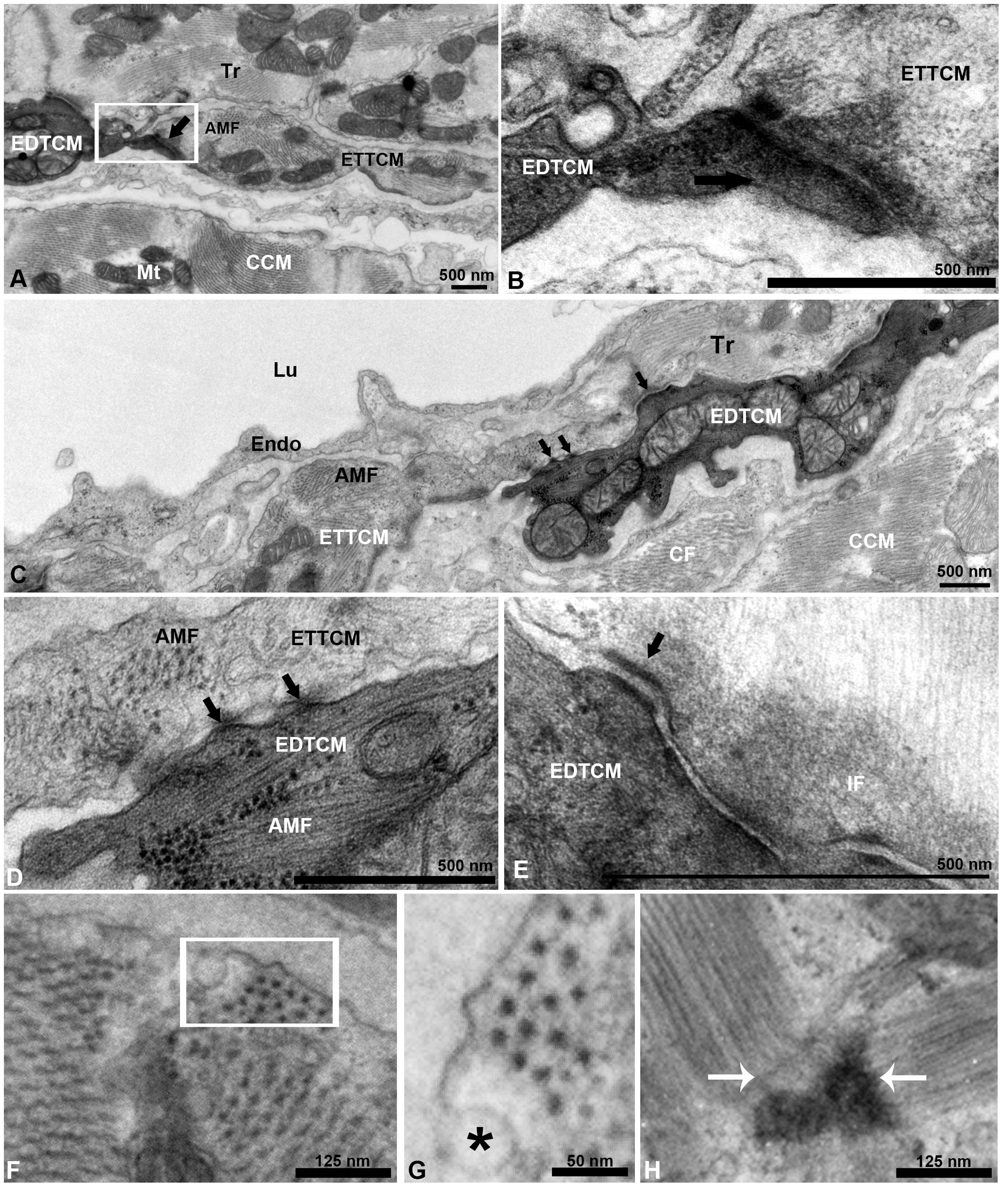
Fig. 2
Electron dense and electron translucent transitional cardiac myocytes interactions.
(A), example of end-to-end interaction between EDTCM and electron translucent cardiac myocytes (ETTCM) forming a continuum at the spongy-compact interface. (B), higher magnification of contact area with complex adhesion junctions including desmosomes between the two cells. (C), example of interaction between mitochondria filled and actin-myosin filament (AMF) containing EDTCM cells and ETTCM via adhesion junctions (arrows). (D), higher magnification of EDTCM and ETTCM showing gap junctions (arrows), and (E), desmosomes. (F), AMF in a transitional cell. (G), higher magnification (inset in F, rotated) showing the hexagonal array of myosin in cardiac myocytes. (F) sarcomere and z-band in the same cell. AMF, actin-myosin filament; CCM, compact cardiac myocytes, CM, cardiac myocytes; EDTCM, electron dense transitional cardiac myocytes; ETTCM, electron translucent transitional cardiac myocytes; Endo, endocardium; Epi; IF, intermediate filaments; Lu, lumen; Mt, mitochondrion; Tr, trabeculae.