
|
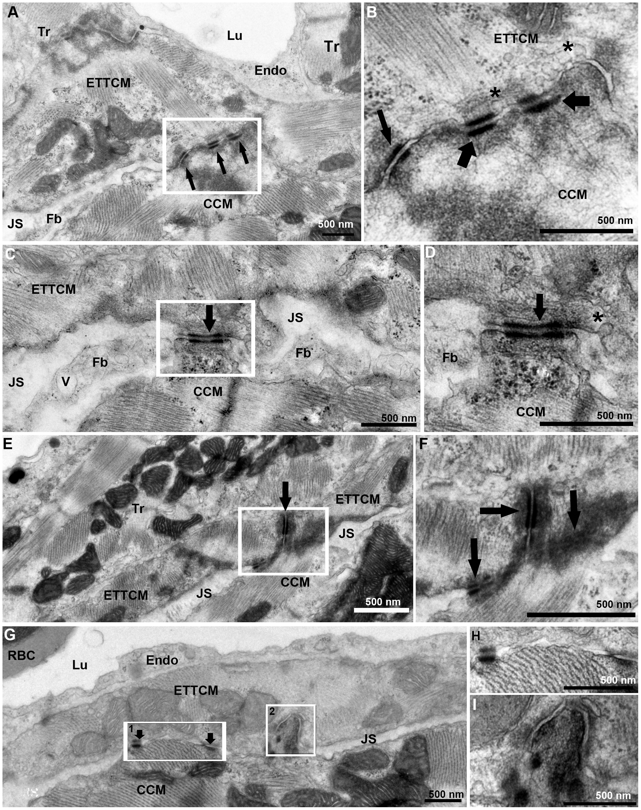
Fig. 6
Transitional CM ring and compact CM contacts.
(A), ETTCM and Compact CM direct contacts mediated by adhesion junctions (arrows). Note the JS with fibroblast (Fb) on the right and narrow JS of the left of the contact region. (B), higher magnification of inset in (A) showing adhesion junctions associated with fascia adherentes (thick arrow), and also with desmosomes (thin arrow). Note the abundance of caveolae (*) in the transitional CM membrane in the contact region. (C), another narrow region of contact (arrow) between a transitional and a compact cardiac myocyte forming a bridge between the two cells and adjacent Z-band in the CCM. A fibroblast process is seen on each side of the junction. Note a vesicle (V) in the fibroblast process. (D), higher magnification of the contact area (arrow) showing a desmosome associated with actin, myosin, and intermediate filaments on the ETTCM and abundant ribosomes (rb) in the CCM. (E), a tri-cellular junction between two transitional cardiac CM and one compact CM. (F), higher magnification of (E, inset) showing desmosomes between the ETTCM and each ETTCM with the compact CM (arrows). (G), discrete adhesion junction between a transitional and compact CM (arrows). (H), higher magnification of inset 1 of (F). (I), higher magnification of inset 2 of F, showing compact CM contact within an invagination of the transitional CM.