
FIGURE
Fig. S3
Fig. S3
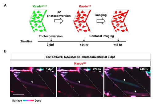
col1a2+ cells contribute to muscle growth. (A) Schematics of photoconversionbased lineage tracing. (B) col1a2Kaede embryos were photoconverted at 3 dpf, and imaged at indicated time points. Color coded depth projections (green corresponds to superficial slices, while red denotes deep slices) of converted Kaedered showed that new Kaedered muscle fibers (arrow) emerged at 48-hour post conversion. An existing muscle fiber present through all 3 time points is indicated by arrowheads. n = 15 embryos. Scale bar: 50 μm. |
Expression Data
Expression Detail
Antibody Labeling
Phenotype Data
Phenotype Detail
Acknowledgments
This image is the copyrighted work of the attributed author or publisher, and
ZFIN has permission only to display this image to its users.
Additional permissions should be obtained from the applicable author or publisher of the image.
Full text @ Development