
Fig. 7
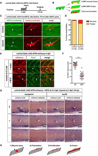
col1a2+ MPCs generate new myofibers primarily by cell fusion. (A) Experimental design. col1a2Cre-ERT2; ubi:Switch embryos treated with 4-OHT for 3.5 h at 3 dpf were either needle injured or left uninjured (controls) and imaged 75 h later. (B) Three possible modes of new myofiber formation. (C) In controls (n=20), new myofibers were formed primarily through cell fusion (arrows), whereas in injured embryos (n=19), new fibers were generated by both fusion (arrows) and occasionally, de novo fiber formation (arrowheads). (D) Quantification of modes of new myofiber formation. The number of new myofibers scored is shown. (E) col1a2NTR-mCherryembryos treated with either water or MTZ at 2-3 dpf were stained with the mCherry (red) and the Pax7 (green) antibodies at 4 dpf. Water-treated controls showed many mCherry+Pax7+ MPCs (arrows), whereas MTZ-treated embryos lost most mCherry+ MPCs with a few spared myofibers (arrowheads). (F) Quantification of the average number of Pax7+ MPCs per somite, including superficial MPCs and deep myofiber-associated MPCs shown in E. n=26 (control) and 25 (MTZ). Data are plotted as mean±s.e.m. Statistics: Mann–Whitney U-test. ****P<0.0001. (G) col1a2NTR-mCherry embryos (ablated) or mCherry− siblings (control) were treated with MTZ at 2-3 dpf, injured at 3 dpf and stained for myogenin or myoD expression at 24 hpi. Embryos were scored based on the extent of expression of myogenic markers at the injury site: high level (++), mid-level (+) and no induction (−). The number of embryos in each category is indicated. Arrows denote induced marker expression and asterisks indicate the injury site in the case of no induction. (H) Model of muscle regeneration by col1a2+ MPCs. Scale bars: 50 µm. |
| Fish: | |
|---|---|
| Condition: | |
| Observed In: | |
| Stage: | Day 4 |