
FIGURE
Fig. S1
Fig. S1
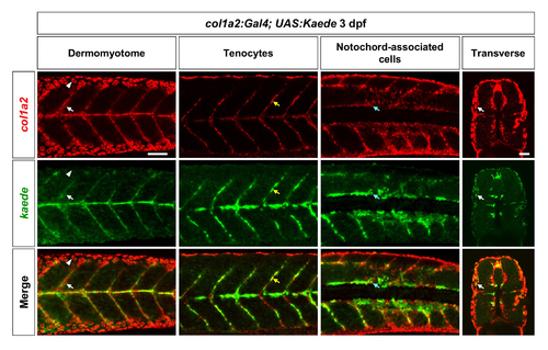
Validation of the col1a2Kaede line. Double fluorescent in situ hybridization using kaede and col1a2 probes were performed in col1a2Kaede embryos at 3 dpf. Co-expression of kaede (green) and the endogenous col1a2 (red) can be observed in dermomyotome cells (white arrows), tenocytes along the vertical myoseptum (yellow arrows), and deep interstitial cells around the notochord (cyan arrows). Note that col1a2Kaede was not expressed in the epidermis (arrowheads) as col1a2. n = 19 embryos. Scale bar: 50 μm. |
Expression Data
Expression Detail
Antibody Labeling
Phenotype Data
Phenotype Detail
Acknowledgments
This image is the copyrighted work of the attributed author or publisher, and
ZFIN has permission only to display this image to its users.
Additional permissions should be obtained from the applicable author or publisher of the image.
Full text @ Development