
Fig. S9
- ID
- ZDB-FIG-170516-36
- Publication
- Münch et al., 2017 - Notch signalling restricts inflammation and serpine1 expression in the dynamic endocardium of the regenerating zebrafish heart.
- Other Figures
- All Figure Page
- Back to All Figure Page
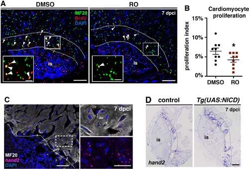
Notch inhibition decreases cardiomyocyte proliferation (A) IHC against BrdU and Mef2 on heart sections (7 dpci), showing lower BrdU incorporation in Mef2+ cells (white arrowheads) adjacent to the injury (between the dashed lines) after treatment with RO than after treatment with DMSO. (B) Scatter plot showing quantification of BrdU+ Mef2+ cells in DMSO- and RO-treated hearts (discontinuous line = mean ± s.d., t-test, *P<0.05). (C) FISH against hand2 combined with IHC for mf20 showing, hand2-expression in cardiomyocytes. (D) ISH against hand2 at 7 dpci, showing higher numbers of injury-adjacent cardiomyocytes expressing these genes (between the dotted lines) in Tg(UAS:NICD) hearts than in control hearts. Dotted lines delineate the injury site (is). Scale bars: 100 µm; 25 µm in amplified views. |