
Fig. S3
- ID
- ZDB-FIG-170516-30
- Publication
- Münch et al., 2017 - Notch signalling restricts inflammation and serpine1 expression in the dynamic endocardium of the regenerating zebrafish heart.
- Other Figures
- All Figure Page
- Back to All Figure Page
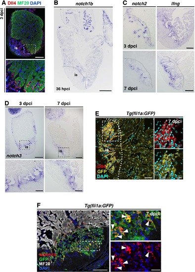
Genes encoding Notch signalling elements are expressed in endocardial cells upon cryoinjury (A) IHC of Dll4 and MF20, showing expression within the injury site (is) and surrounding adjacent cardiomyocytes. The boxed area is magnified in the panel below. (B) ISH for notch1b, showing low notch1b expression in the injury site and the remote region at 36 hpci. The dotted line demarcates the injury site (is). (C, D) ISH of lfng, notch2 and notch3 in regenerating hearts, showing expression adjacent to and within the injury site (is; 3 dpci, 7 dpci). Boxed areas are shown at higher magnification in the lower row. (E) IHC against Dll4 and GFP on sections of Tg(fli1a:GFP) transgenic hearts (7 dpci), showing endocardial expression of Dll4 (white arrowheads). The dotted line demarcates the injury site (is). (F) FISH against notch1b combined with IHC against GFP and MF20 on sections of Tg(fli1a:GFP) transgenic hearts (7 dpci) showing notch1b transcripts in endocardial cells (white arrowheads). The boxed area is magnified in the right-hand panels. The dotted line demarcates the injury site (is). (Scale bars: 200 µm in A, B; 100 µm in C, D, E, F; 50 µm in magnified views in A, D; 20 µm in E, F. |
| Genes: | |
|---|---|
| Fish: | |
| Condition: | |
| Anatomical Term: | |
| Stage: | Adult |