
Fig. S6
- ID
- ZDB-FIG-170516-33
- Publication
- Münch et al., 2017 - Notch signalling restricts inflammation and serpine1 expression in the dynamic endocardium of the regenerating zebrafish heart.
- Other Figures
- All Figure Page
- Back to All Figure Page
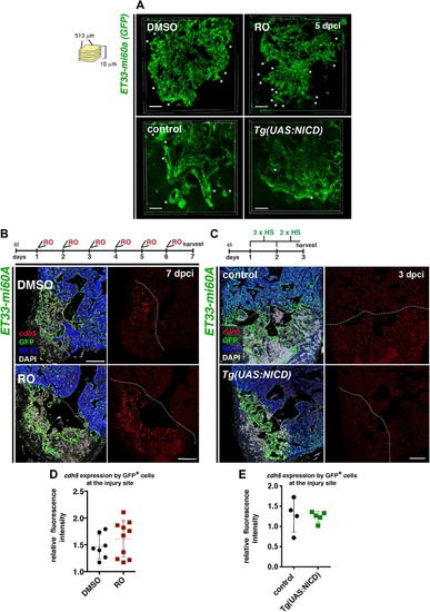
Notch signalling modulation does not interfere with endocardial cdh5-expression (A) Volume rendering of part of the injured region (513 μm x 513 μm x 10 μm) from ET33mi-60A; myl7mRFP hearts after treatment with RO or DMSO and from ET33mi-60A; myl7mRFP, Tg(UAS:NICD) and control hearts. Comparable 3D-images were used for quantification of filopodia- like protrusions. The graph of quantified filopodia is shown in Figure 3I. (B, C) FISH against cdh5 combined with IHC against GFP and mf20 on sections of ET33-mi60a transgenic hearts after treatment with DMOS or RO (7dpci) and of ET33mi-60A;Tg(UAS:NICD) and control hearts ( 3dpci). The treatment regime is indicated on top. GFP+ endocardial/endothelial cells at the injury site present similar levels of cdh5 expression in injured hearts after treatment with RO or DMSO and in ET33mi-60A;Tg(UAS:NICD) or control hearts. (D, E) Scatter plot showing relative red fluorescence intensity, comparing values of the endocardium of the remote region to the injury endocardium (see Figure 2D), in injured hearts (dotted line = mean± s.d, t-test, not significant). Scale bars: 50 μm in A, 100 μm in B, C. |