
Fig. 1
- ID
- ZDB-FIG-170516-21
- Publication
- Münch et al., 2017 - Notch signalling restricts inflammation and serpine1 expression in the dynamic endocardium of the regenerating zebrafish heart.
- Other Figures
- All Figure Page
- Back to All Figure Page
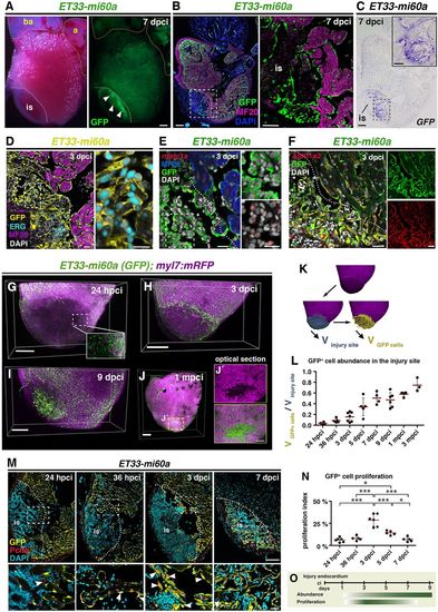
The endocardium expands at the injury site. (A) ET33-mi60a transgenic zebrafish heart, whole-mount views. Strong GFP expression is apparent in the cryoinjured region of the ventricle (arrowheads). (B) ET33-mi60a heart section. Immunohistochemistry (IHC) shows GFP+ endocardial cells in a myosin heavy chain (MF20)− area. (C) ET33-mi60a heart consecutive section to Fig. 1B showing GFP ISH. (D-F) IHC, or IHC combined with FISH. GFP+ cells express Erg (D), nfatc1a (E) and Aldh1a2 (F). (B-F) Boxed areas are magnified on the right. (G-I) Volume rendering of ET33-mi60a;myl7:mRFP injured ventricles. Endocardium, green; myocardium, magenta. (J,J′) Volume rendering (J) and amplified optical section from the centre (J′) of an injured ventricle. (K) Imaris-based volume quantification. The volume of the mRFP− region was determined (blue, Vinjury site) and used as a mask to label GFP+ cells in this region (yellow) and determine their volume (VGFP cells). (L) Scatter plot showing the relative volume occupied by GFP+ cells in the injury site. Mean (red line)±s.d.; one-way ANOVA and Newman-Keuls test (see Table S3). (M) IHC showing Pcna+ GFP+ endocardial nuclei (arrowheads). Boxed areas magnified beneath. (N) Scatter plot showing percentage of Pcna+ cells among GFP+ cells within and adjacent (50 μm) to the injury site. Mean±s.d.; one-way ANOVA and Newman-Keuls test, *P<0.05, ***P<0.005 (see Table S4). (O) Schematic illustrating that endocardium proliferation precedes endocardial expansion at the injury site. is, injury site; a, atrium; ba, bulbus arterious. Dotted lines demarcate injured tissue. Scale bars: 200 μm in A-C,G-J; 20 μm in D-F; 100 μm in M; 50 μm in magnified views in B,C; 20 μm in magnified views in D,E,M; 100 μm in J. |