
Fig. S3
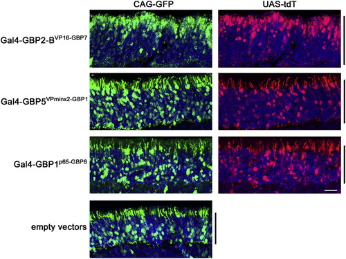
T-DDOGS Composed of VPminx2AD and p65AD Give Normal Positioning of Rod Photoreceptors in the Retina, Related to Figure 4 Representative images showing the effects of selected T-DDOGs on rod photoreceptor positioning. CD1 mouse retinas were electroporated at P0 in vivo with plasmids encoding CAG-GFP (green), Gal4-GBPXAD-GBPY components, UAS-tdT (red) and CAG-nlacZ. The electroporated GFP:DBDG:ADG plasmid mass ratio is 2:2:1. Retinas were harvested at P14. Confocal stacks of retina sections spanning the ONL (black bar) are shown. Each set of panels was adjusted individually to similar levels to reveal the cell bodies. DAPI is blue in all panels. Scale bar, 20 μm. Non-linear adjustment using the gamma function was applied to show weak GFP or tdT-expressing cells to minimize their signals from being obscured by those of stronger-expressing equivalents. The scleral portion of the ONL is the upper half of each image. |
Reprinted from Cell, 154(4), Tang, J.C., Szikra, T., Kozorovitskiy, Y., Teixiera, M., Sabatini, B.L., Roska, B., and Cepko, C.L., A nanobody-based system using fluorescent proteins as scaffolds for cell-specific gene manipulation, 928-939, Copyright (2013) with permission from Elsevier. Full text @ Cell