
FIGURE
Fig. S1
Fig. S1
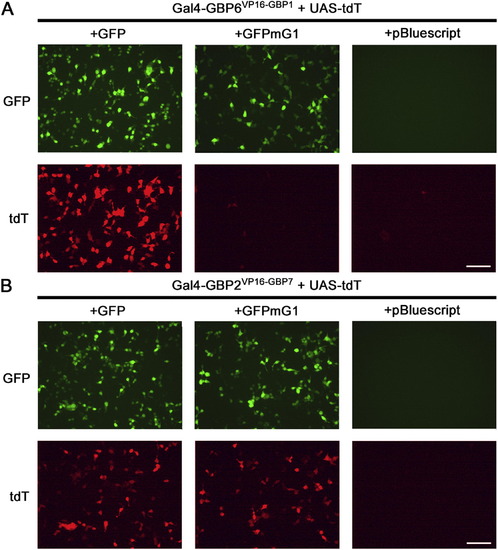
GFP-Dependent Activation of UAS-tdT In Vitro, Related to Figure 2 (A and B) Representative images of Gal4-GBP6VP16-GBP1 (A) and Gal4-GBP2VP16-GBP7 (B) activation of an UAS-tdT reporter in 293T cells. Cells were transfected with all indicated constructs and imaged 16 hr post-transfection. GFP, GFPmG1, DBDG and ADG constructs were delivered in separate plasmids under the CAG-promoter. Results are representative of 4 independent experiments and quantifications are tabulated in Table S2. Scale bar, 100 μm. |
Expression Data
Expression Detail
Antibody Labeling
Phenotype Data
Phenotype Detail
Acknowledgments
This image is the copyrighted work of the attributed author or publisher, and
ZFIN has permission only to display this image to its users.
Additional permissions should be obtained from the applicable author or publisher of the image.
Reprinted from Cell, 154(4), Tang, J.C., Szikra, T., Kozorovitskiy, Y., Teixiera, M., Sabatini, B.L., Roska, B., and Cepko, C.L., A nanobody-based system using fluorescent proteins as scaffolds for cell-specific gene manipulation, 928-939, Copyright (2013) with permission from Elsevier. Full text @ Cell