
FIGURE
Fig. S2
Fig. S2
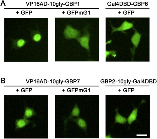
VP16AD-GBP Fusions Can Localize GFP to the Nucleus, Related to Figure 2 Representative images of GFP localization in 293T cells transfected with pCAG-GFP or pCAG-GFPmG1 along with CAG-driven DBDG or ADG plasmids at a 1:2 molar plasmid ratio. Fluorescent micrographs were taken at 16 hr post-transfection in live cells. Scale bar, 15μm. |
Expression Data
Expression Detail
Antibody Labeling
Phenotype Data
Phenotype Detail
Acknowledgments
This image is the copyrighted work of the attributed author or publisher, and
ZFIN has permission only to display this image to its users.
Additional permissions should be obtained from the applicable author or publisher of the image.
Reprinted from Cell, 154(4), Tang, J.C., Szikra, T., Kozorovitskiy, Y., Teixiera, M., Sabatini, B.L., Roska, B., and Cepko, C.L., A nanobody-based system using fluorescent proteins as scaffolds for cell-specific gene manipulation, 928-939, Copyright (2013) with permission from Elsevier. Full text @ Cell