
- Title
-
Upregulation of the zebrafish Nogo-A homologue, Rtn4b, in retinal ganglion cells is functionally involved in axon regeneration
- Authors
- Welte, C., Engel, S., Stuermer, C.A.
- Source
- Full text @ Neural Dev.
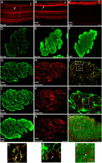
Expression pattern of Rtn4b in the zebrafish retina and optic nerve. Cross sections of the zebrafish retina normal (A) and 10 days after ONS (B,C) were exposed to ABs against Rtn4b (A,B) and MBP (C). Weak Rtn4b staining is seen across all retinal layers including RGCs (white arrow) in the normal retina (A). RGCs robustly upregulate Rtn4b 10 days after ONS (B). The RGC axons in the retina on top of the RGCs (bracket) are also weakly labeled but are more intensely stained by the AB against MBP (C). Scale bar, 50 µm. Cross sections through the normal zebrafish optic nerve (D,E,F) show very weak labeling with Rtn4b AB (D). (E) Rtn4a AB stains across the normal nerve similar to MBP. Labeling with the MBP AB is strong in the normal nerve (F) as well as at 10 days after ONS (M). Scale bar, 10 µm. Rtn4b AB in nerves at 10 days after ONS (G,I) labels portions of RGC axons identified as axonal (rather than glial) by the AB against neurolin (H). (I), overlay. Scale bar, 20 µm. (J) Rtn4a AB in the10-days ONS nerve stains, in particular, the boundaries of fascicles and subdivisions of the fascicles. (K,L) The neurolin-positive regenerating RGC axons are not labeled with Rtn4a to any significant extent, in contrast to Rtn4b labeling (I). (M) MBP staining is intense in the 10-day ONS nerve. (N,O) Neurolin-positive regenerating RGC axons are located amidst the myelin. Boxed areas in (I,L,O) are enlarged in (P,Q,R). Arrows point to neurolin-positive axons which are also Rtn4b AB-positive (P), but seem not significantly co-localized with Rtn4a (Q), and lie amidst the MBP labeling of myelin (R). Scale bar, 10 µm. EXPRESSION / LABELING:
|
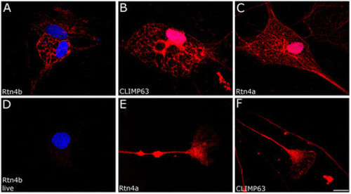
Rtn4a and Rtn4b staining of ER in zebrafish oligodendrocytes. (A) Labeling of fixed zebrafish oligodendrocytes with Rtn4b reveals a reticular structure similar to the ER staining with CLIMP63 AB (B). (C) Rtn4a AB also labels ER in oligodendrocytes. (D) Exposing live oligodendrocytes to Rtn4b AB gives no cell surface staining. DAPI stains the nuclei in (A,D) and also in (B,C) where, however, the red ER stain covers the blue. (E) Zebrafish RGC axons and growth cones are labeled by the Rtn4b AB. These structures are also labeled with CLIMP63 AB (F). Scale bar, 10 µm. EXPRESSION / LABELING:
|
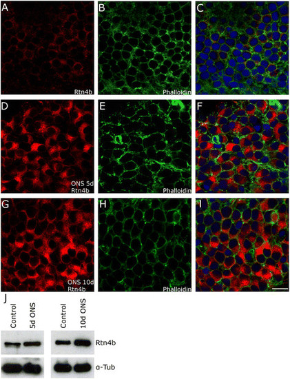
Upregulation of Rtn4b in zebrafish RGCs after ONS. RGCs in retina whole mounts showed weak immunostainings in the cytoplasm after exposure to Rtn4b AB (A). (B,E,H) Labeling with Phalloidin against F-actin shows all cells and their cytoplasm. (C,F,I) Merge of (A,B), (C,D), and (G,H) with DAPI stainings to visualize nuclei. (D,E,F) 5 days after ONS, the RGCs exhibit a significant increase in size and increase in Rtn4b labeling intensity (48% in comparison to control, P < 0.01) in the cytoplasm. (G,H,I) 10 days after optic nerve sections, the size on the RGCs and the intensity of Rtn4b staining is still highly increased (53% over controls, P < 0.01). Scale bar, 10 µm. (J) This apparent increase in the 100 kd Rtn4b protein is also seen in Western blots with retinae at 5 (P < 0.05) and 10 days (P < 0.0001, Student’s T-test) after ONS. Anti-alpha tubulin served as loading control. EXPRESSION / LABELING:
|
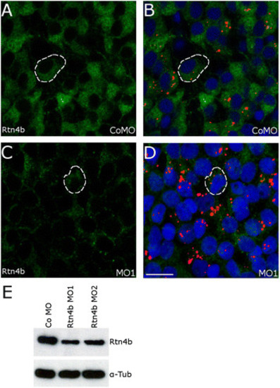
Downregulation of Rtn4b in RGCs by MO1. After application of the control (Co) MO to the lesioned optic nerve, Rtn4b labeling of RGCs in retina whole mounts was intense at 5 days after ONS (A,B), but was markedly reduced when MO1 against Rtn4b (C,D) was offered. (B,D) The Rtn4b AB-labeled cells contain Lissamine (red) conjugated to the MOs. Examples of RGCs are outlined (white interrupted lines). DAPI stains the nuclei. Scale bar, 10 µm. (E) Western blot analysis showing a significant decrease in Rtn4b expression in retinae 5 days after ONS and application of MO1 and MO2 (P < 0.0001), respectively, to the optic nerve. Alpha-tubulin served as loading control. EXPRESSION / LABELING:
|
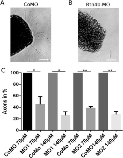
Quantification of axon number after MO application in the outgrowth assay. (A,B) Retina mini-explants isolated from retinae 5 days after (A) control MO (Co) or (B) Rtn4b MO1 application to the optic nerve, extend axons and axon fascicles (arrows) after 24 h in vitro. Outgrowth is significantly reduced on the Rtn4b MO-treated side. Scale bar, 100 µm. (C) The histogram demonstrates the decline in the number of axons extending from retina explants in vitro after MO1 and MO2 application to the optic nerve, in comparison to axon number from control (Co) MO-treated fish (100%). Bars indicate standard deviation. The differences between groups are statistically significantly different. Quantification was done on three replicates from three different experiments, and for statistical analysis, Student’s T-test was used. *P < 0.05, **P < 0.01. PHENOTYPE:
|
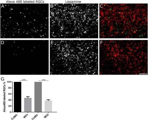
Rtn4b MO-induced reduction in axon regeneration in the in vivo regeneration assays. (A-F) After application of Alexa488 to the regenerating axons (distal from the original lesion and MO application site), the retrogradely labeled RGCs are counted in retina whole mounts. Many more Alexa488-labeled RGCs are recognized 9 days after ONS and control (Co) MO application (A) than on the contralateral retina (D) belonging to the nerve that received Rtn4b MO1 (or MO2). (B, E) The RGCs contain lissamine associated with the MOs. (C,F) Merge of (A,B) and (D,E). Scale bar, 50 µm. (G) The histogram demonstrates the decline in the number of Alexa-labeled RGCs after MO1 and MO2 application to the optic nerve, in comparison to axon number from control (Co) MO-treated fish (100%). Bars indicate standard deviation. Three different experiments with n, 10 retinal squares (300 × 300 µm) for each experimental group were statistically evaluated using Student’s T-test. The differences between groups are statistically significantly different, ***P < 0.001. PHENOTYPE:
|