
Fig. 6
- ID
- ZDB-FIG-150806-2
- Publication
- Welte et al., 2015 - Upregulation of the zebrafish Nogo-A homologue, Rtn4b, in retinal ganglion cells is functionally involved in axon regeneration
- Other Figures
- All Figure Page
- Back to All Figure Page
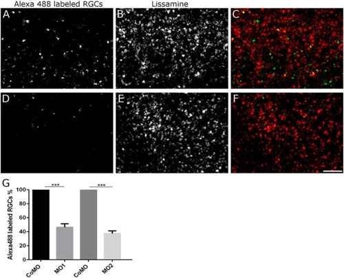
Rtn4b MO-induced reduction in axon regeneration in the in vivo regeneration assays. (A-F) After application of Alexa488 to the regenerating axons (distal from the original lesion and MO application site), the retrogradely labeled RGCs are counted in retina whole mounts. Many more Alexa488-labeled RGCs are recognized 9 days after ONS and control (Co) MO application (A) than on the contralateral retina (D) belonging to the nerve that received Rtn4b MO1 (or MO2). (B, E) The RGCs contain lissamine associated with the MOs. (C,F) Merge of (A,B) and (D,E). Scale bar, 50 µm. (G) The histogram demonstrates the decline in the number of Alexa-labeled RGCs after MO1 and MO2 application to the optic nerve, in comparison to axon number from control (Co) MO-treated fish (100%). Bars indicate standard deviation. Three different experiments with n, 10 retinal squares (300 × 300 µm) for each experimental group were statistically evaluated using Student’s T-test. The differences between groups are statistically significantly different, ***P < 0.001. |
| Fish: | |
|---|---|
| Condition: | |
| Knockdown Reagents: | |
| Observed In: | |
| Stage: | Adult |