
Fig. 3
- ID
- ZDB-FIG-150528-22
- Publication
- Welte et al., 2015 - Upregulation of the zebrafish Nogo-A homologue, Rtn4b, in retinal ganglion cells is functionally involved in axon regeneration
- Other Figures
- All Figure Page
- Back to All Figure Page
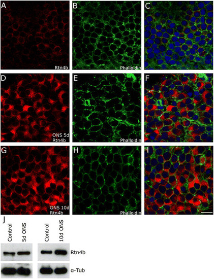
Upregulation of Rtn4b in zebrafish RGCs after ONS. RGCs in retina whole mounts showed weak immunostainings in the cytoplasm after exposure to Rtn4b AB (A). (B,E,H) Labeling with Phalloidin against F-actin shows all cells and their cytoplasm. (C,F,I) Merge of (A,B), (C,D), and (G,H) with DAPI stainings to visualize nuclei. (D,E,F) 5 days after ONS, the RGCs exhibit a significant increase in size and increase in Rtn4b labeling intensity (48% in comparison to control, P < 0.01) in the cytoplasm. (G,H,I) 10 days after optic nerve sections, the size on the RGCs and the intensity of Rtn4b staining is still highly increased (53% over controls, P < 0.01). Scale bar, 10 µm. (J) This apparent increase in the 100 kd Rtn4b protein is also seen in Western blots with retinae at 5 (P < 0.05) and 10 days (P < 0.0001, Student’s T-test) after ONS. Anti-alpha tubulin served as loading control. |
| Gene: | |
|---|---|
| Antibody: | |
| Fish: | |
| Condition: | |
| Anatomical Terms: | |
| Stage: | Adult |