
- Title
-
The voltage-gated calcium channel CACNB2 (β2.1) protein is required in the heart for control of cell proliferation and heart tube integrity
- Authors
- Chernyavskaya, Y., Ebert, A.M., Milligan, E., and Garrity, D.M.
- Source
- Full text @ Dev. Dyn.
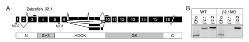
β2.1 gene structure and morpholino (MO). A: Exon map of zebrafish β2.1 protein showing sites of alternative splicing and MAGUK domains. The SH3 and GK domains (gray) are conserved in all MAGUK proteins. The MO1 binding site spans the splice donor sequences located at the 3′ end of exon 5, and primers used for reverse transcriptasepolymerase chain reaction (RTPCR) are indicated in exons 3 and 10. The MO2 binding site spans the ATG start site located in exon 1. B: RTPCR to assess the efficacy of the β2.1 MO1 in reducing fulllength β2.1 mRNA. In RNA from a wildtype embryo (left), β2.1 and β2.2 transcripts (each detected with genespecific primers) are present at 48 hours postfertilization (hpf). In RNA extracted from 48 hpf embryos injected with 750 μM β2.1 MO1 (right), β2.1 transcripts were not detectable, whereas expression of β2.2 transcript levels were indistinguishable from wildtype. The resulting amplicons are 200 bp (ef1α), 428 bp (β2.1), and 467 bp (β2.2). EXPRESSION / LABELING:
|
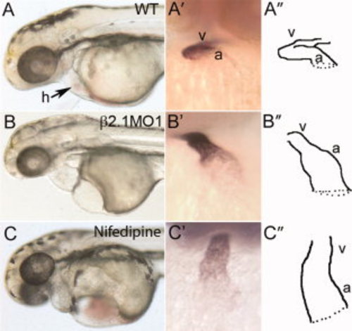
Cardiac phenotypes in β2.1-depleted or LTCC-inhibited embryos. A–C: Brightfield images of 72 hours postfertilization (hpf) embryos injected with buffer only (A), or β2.1-MO1 (B), or exposed to 20 μM nifedipine (an LTCC antagonist, C) for 24 hr. A2–C2) In situ hybridization showing hearts of 48 hpf embryos labeled with a probe against myl-7. A3–C3) Tracings of 48 hpf hearts to delineate chamber morphology. MO, morpholino; a, atrium; h, heart; v, ventricle. EXPRESSION / LABELING:
PHENOTYPE:
|
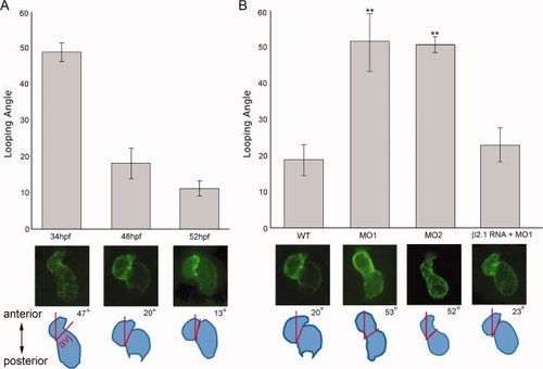
Quantification of looping using plane of the atrioventricular junction to define the “looping angle.” A: Wild-type Tg(myl7:EGFP HsHRAS) hearts were imaged. The “looping angle” was defined as the angle created between the plane of the cardiac atrioventricular junction and the embryo anteroposterior axis, as diagrammed. A: In wild-type embryos, the average looping angle decreased significantly as development proceeded (P < 0.001; n = 20 embryos/time point). B: At 48 hpf, MO1 or MO2 morphant hearts displayed a significantly greater average looping angle than wild-type (P < 0.01; n = 20 embryos/time point), indicating less looping had occurred. The looping angle was restored to wild-type ranges by co-injecting β2.1cRNA along with MO1 (P = 0.40). avj, atrioventricular junction. |
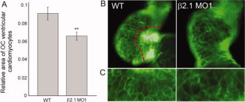
Cell morphology in wild-type and β2.1-depleted cardiomyocytes. A: Relative area of cells found in the outer curvature (OC) of the wild-type (n = 40) or β2.1-depleted hearts (n = 36) at 48 hours postfertilization (hpf). Asterisks denote the significant difference (P = 0.003). B: Tg(myl7:EGFP-HsHRAS) hearts were imaged as the basis for calculating cell area in OC cells. OC cells lie to the left of the red delineation. C: A magnified view of representative OC cells (from photos in B, rotated 90°) showing the elongated shape of wild-type cells compared with the rounder shape of β2.1-depleted cells. |
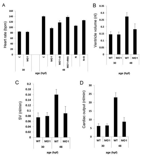
Physiological studies on heart function in β2.1-depleted embryos. A: Average heart rates at 30 and 48 hours postfertilization (hpf) for embryos were measured in beats per min (bpm). Where noted, embryos were exposed to combinations of 20 μM nifedipine (an LTCC antagonist) for 24 hr (from 24 to 48 hpf), 40 μM BayK (an LTCC agonist) for ~30 min (from 47.5 to 48 hpf) or β2.1 morpholino (injected at the 1-4-cell stage). B–D: The volume of the ventricle at diastole (A), ventricular stroke volume (C), and cardiac output (D) were calculated from selected frames of videos recording ventricular contraction. n = 9 embryos per treatment. B, BayK; C, control; MO, β2.1 morpholino MO1; N, nifedipine. |
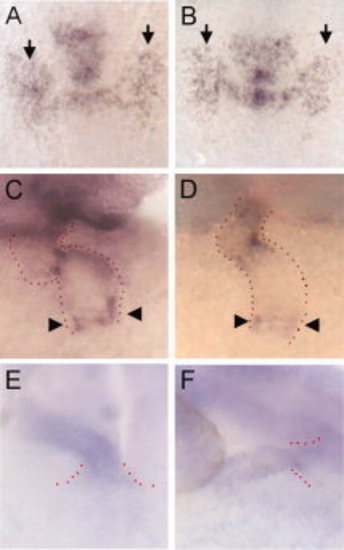
Differentiation of cells in the first heart field and at the venous and arterial poles. A,B:fgf8 expression in the bilateral heart fields (arrows) at the four-somite stage (11.5 hours postfertilization [hpf]) is comparable in wild-type and morphant embryos. C,D: In situ hybridization with bmp4 on 48 hpf embryos, indicating normal expression of this marker at the venous end of the heart tube (arrowheads). Red dots outline the shape of the heart tube. E,F: In situ hybridization with vhmc on 30 hpf embryos, indicating the presumptive ventricle, as well as secondary heart field cells being added to the arterial pole (flanked by red dots). EXPRESSION / LABELING:
PHENOTYPE:
|

Chamber-specific cell counts at 48 hours postfertilization (hpf). Cardiomyocyte populations in wild-type and β2.1-depleted heart chambers at 48 hpf, based on counts of dsRED-labeled nuclei (n = 20 embryos/treatment). Significance is designated by asterisks (P = 0.02). PHENOTYPE:
|
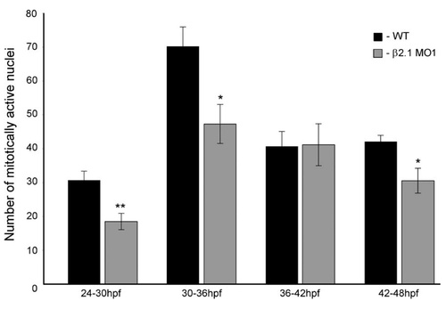
Bromodeoxyuridine (BrdU) assay of cardiomyocyte cells undergoing DNA replication, spanning 6-hr periods of cardiac development. β2.1-depleted embryos display a significantly reduced rate of mitosis (asterisks) at 24–30 hpf (P = 0.002), 30–36 hpf (P = 0.01), and 42–48 hpf (P = 0.01). n = 10 wild-type and 10 morphant embryos per time period. |
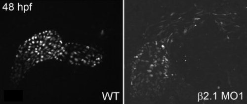
Adhesion assays to examine heart tube integrity in wild-type vs. β2.1-depleted heart tubes. At 48 hours postfertilization (hpf), Tg(myl7:nDsRed2) embryos were subjected to slight pressure from a coverslip to determine how well the heart was able to withstand stress. β2.1 hearts dissociated more easily than wild-type heart subjected to the same pressure (n = 20 embryos/treatment). |
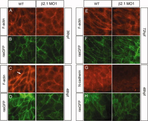
Integrity of cytoskeletal structures in wild-type and β2.1-depleted ventricular cardiomyocytes. A–F: A time course of cardiomyocyte cytoskeletal structure is presented, with hearts at 36 hpf (A,B), 48 hpf (C,D), and 72 hpf (E,F). Wild-type cardiomyocytes contain cytoskeletal actin fibers that localize near the cell membrane (indicated by green fluorescent protein [GFP] signal in B,D,F and H). Some of the actin present is condensed and arranged within the sarcomeres (arrow). Morphant cardiomyocytes still show the presence of cytoskeletal actin at the membrane, but the presence of sarcomeric actin is not as prevalent. G,H: Wild-type cells maintain intra-cellular cohesion by expressing N-cadherin at the membrane while β2.1-depleted cells lack substantial N-cadherin expression at their cell membranes. EXPRESSION / LABELING:
PHENOTYPE:
|
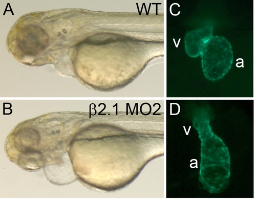
Cardiac phenotypes in morpholino (MO) 2 morphant embryos. A,B: Brightfield images of 72 hours postfertilization (hpf) embryos injected with buffer only (A), or β2.1-MO2 (B). C,D: Hearts of 48 hpf Tg(myl7:nDsRed2) embryos injected with buffer only (C), or β2.1-MO2 (D). |