|
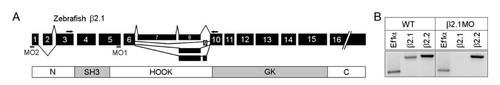
β2.1 gene structure and morpholino (MO). A: Exon map of zebrafish β2.1 protein showing sites of alternative splicing and MAGUK domains. The SH3 and GK domains (gray) are conserved in all MAGUK proteins. The MO1 binding site spans the splice donor sequences located at the 3′ end of exon 5, and primers used for reverse transcriptasepolymerase chain reaction (RTPCR) are indicated in exons 3 and 10. The MO2 binding site spans the ATG start site located in exon 1. B: RTPCR to assess the efficacy of the β2.1 MO1 in reducing fulllength β2.1 mRNA. In RNA from a wildtype embryo (left), β2.1 and β2.2 transcripts (each detected with genespecific primers) are present at 48 hours postfertilization (hpf). In RNA extracted from 48 hpf embryos injected with 750 μM β2.1 MO1 (right), β2.1 transcripts were not detectable, whereas expression of β2.2 transcript levels were indistinguishable from wildtype. The resulting amplicons are 200 bp (ef1α), 428 bp (β2.1), and 467 bp (β2.2).
|