
|
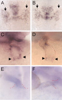
Fig. 6
Differentiation of cells in the first heart field and at the venous and arterial poles. A,B:fgf8 expression in the bilateral heart fields (arrows) at the four-somite stage (11.5 hours postfertilization [hpf]) is comparable in wild-type and morphant embryos. C,D: In situ hybridization with bmp4 on 48 hpf embryos, indicating normal expression of this marker at the venous end of the heart tube (arrowheads). Red dots outline the shape of the heart tube. E,F: In situ hybridization with vhmc on 30 hpf embryos, indicating the presumptive ventricle, as well as secondary heart field cells being added to the arterial pole (flanked by red dots).