
Image
|
Figure Caption
Fig. 2
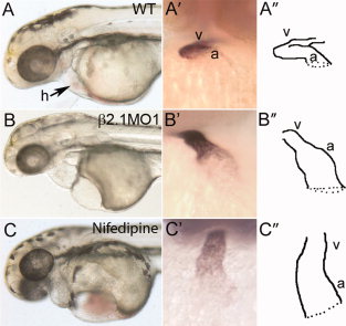
Cardiac phenotypes in β2.1-depleted or LTCC-inhibited embryos. A–C: Brightfield images of 72 hours postfertilization (hpf) embryos injected with buffer only (A), or β2.1-MO1 (B), or exposed to 20 μM nifedipine (an LTCC antagonist, C) for 24 hr. A2–C2) In situ hybridization showing hearts of 48 hpf embryos labeled with a probe against myl-7. A3–C3) Tracings of 48 hpf hearts to delineate chamber morphology. MO, morpholino; a, atrium; h, heart; v, ventricle.
Figure Data
Acknowledgments
This image is the copyrighted work of the attributed author or publisher, and
ZFIN has permission only to display this image to its users.
Additional permissions should be obtained from the applicable author or publisher of the image.
Full text @ Dev. Dyn.