
Image
|
Figure Caption
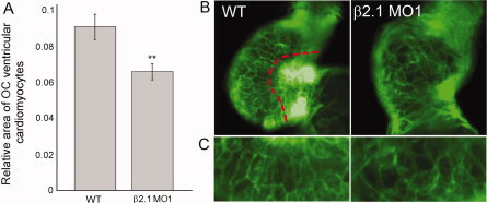
Fig. 4
Cell morphology in wild-type and β2.1-depleted cardiomyocytes. A: Relative area of cells found in the outer curvature (OC) of the wild-type (n = 40) or β2.1-depleted hearts (n = 36) at 48 hours postfertilization (hpf). Asterisks denote the significant difference (P = 0.003). B: Tg(myl7:EGFP-HsHRAS) hearts were imaged as the basis for calculating cell area in OC cells. OC cells lie to the left of the red delineation. C: A magnified view of representative OC cells (from photos in B, rotated 90°) showing the elongated shape of wild-type cells compared with the rounder shape of β2.1-depleted cells.
Figure Data
Acknowledgments
This image is the copyrighted work of the attributed author or publisher, and
ZFIN has permission only to display this image to its users.
Additional permissions should be obtained from the applicable author or publisher of the image.
Full text @ Dev. Dyn.