
|
Fig. 3
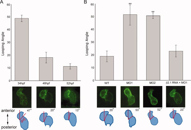
Quantification of looping using plane of the atrioventricular junction to define the “looping angle.” A: Wild-type Tg(myl7:EGFP HsHRAS) hearts were imaged. The “looping angle” was defined as the angle created between the plane of the cardiac atrioventricular junction and the embryo anteroposterior axis, as diagrammed. A: In wild-type embryos, the average looping angle decreased significantly as development proceeded (P < 0.001; n = 20 embryos/time point). B: At 48 hpf, MO1 or MO2 morphant hearts displayed a significantly greater average looping angle than wild-type (P < 0.01; n = 20 embryos/time point), indicating less looping had occurred. The looping angle was restored to wild-type ranges by co-injecting β2.1cRNA along with MO1 (P = 0.40). avj, atrioventricular junction.