
|
Fig. 10
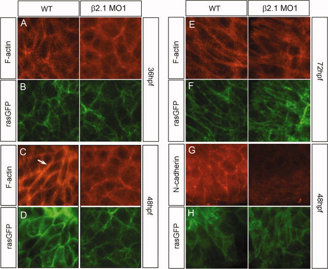
Integrity of cytoskeletal structures in wild-type and β2.1-depleted ventricular cardiomyocytes. A–F: A time course of cardiomyocyte cytoskeletal structure is presented, with hearts at 36 hpf (A,B), 48 hpf (C,D), and 72 hpf (E,F). Wild-type cardiomyocytes contain cytoskeletal actin fibers that localize near the cell membrane (indicated by green fluorescent protein [GFP] signal in B,D,F and H). Some of the actin present is condensed and arranged within the sarcomeres (arrow). Morphant cardiomyocytes still show the presence of cytoskeletal actin at the membrane, but the presence of sarcomeric actin is not as prevalent. G,H: Wild-type cells maintain intra-cellular cohesion by expressing N-cadherin at the membrane while β2.1-depleted cells lack substantial N-cadherin expression at their cell membranes.