
- Title
-
Zebrafish and fly Nkx6 proteins have similar CNS expression patterns and regulate motoneuron formation
- Authors
- Cheesman, S.E., Layden, M.J., Von Ohlen, T., Doe, C.Q., and Eisen, J.S.
- Source
- Full text @ Development
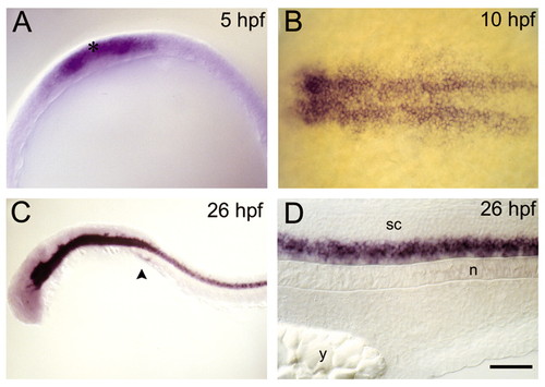
nkx6.1 is expressed in zebrafish ventral spinal cord. (A) Expression is initiated in the embryonic shield at 50% epiboly, approximately 5 hpf (animal pole view, optical section of embryo without yolk). Asterisk denotes epiblast (outer) layer. (B) By bud stage, ∼10 hpf, nkx6.1 is broadly expressed in medial neurectoderm (dorsal view, rostral to left). (C) At 26 hpf, nkx6.1 is strongly expressed in ventral CNS from the midbrain through the tail (lateral view; rostral to left). Arrowhead denotes pancreas expression. (D) Higher magnification view of spinal cord shown in (C). Scale bar: 50 μm for A,B,D; 25 μm for C. sc, spinal cord; n, notochord; y, yolk. EXPRESSION / LABELING:
|
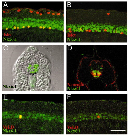
Nkx6.1 is expressed in zebrafish motoneurons. Nkx6.1 (green) Islet1/2 (red) antibody double labels. (A) 14 hpf lateral view reveals Rohon-Beard neurons (RBs) in dorsal spinal cord express only Islet proteins and PMNs in ventral spinal cord are double labeled (yellow). (B) By 18 hpf Nkx6.1 is largely absent from PMNs; most RBs are out of focal plane. (C) Cross-section of 24 hpf embryo showing Nkx6.1-positive nuclei, which comprise about five cell rows in the ventral spinal cord including medial (^) and lateral (*) floorplate. (D) Cross-section of 48 hpf embryo double labeled with Nkx6.1 (green) and Neurolin (red) antibodies. Nkx6.1-positive nuclei are surrounded by Neurolin-labeled cell surfaces. Laterally-located slow muscle cells are also Neurolin-positive. (E) 20 hpf lateral view of individually labeled VeLD interneuron (yellow) expressing Nkx6.1 (green). Most individually labeled VeLDs in 20-22 hpf embryos expressed Nkx6.1 (n=11, 82%). (F) By 24 hpf VeLDs have downregulated Nkx6.1 (n=13, 85%). Scale bar: 50 μm for A,B,D,E,F; 75 μm for C. EXPRESSION / LABELING:
|
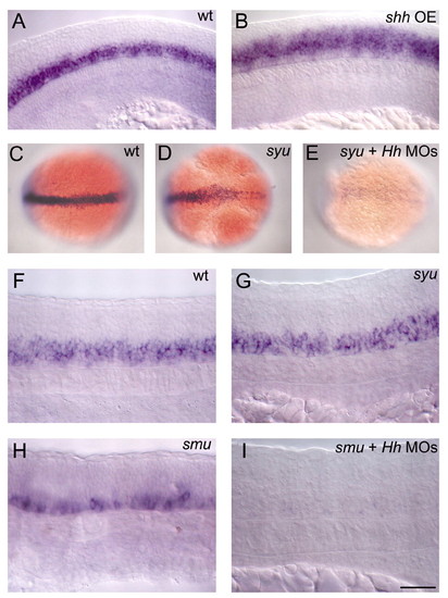
Hh signaling induces zebrafish nkx6.1 expression. (A) 18 hpf wild-type expression of nkx6.1. (B) Dramatic dorsal expansion of nkx6.1 in an 18 hpf embryo ectopically expressing shh (shh OE, 87% of injected animals, n=283). (C) 12 hpf wild-type expression of nkx6.1. (D) 12 hpf syu mutant with reduced nkx6.1 expression. (E) 12 hpf syu mutant injected with ehh and twhh MOs. nkx6.1 expression is largely absent (28% of injected clutch from syu heterozygous parents, n=120). (F) Wild-type expression of nkx6.1 in spinal cord at 24 hpf. (G) 24 hpf syu mutant has wild-type levels of nkx6.1 expression in spinal cord. (H) Few nkx6.1-positive cells remain in a 24 hpf smu mutant spinal cord. (I) 24 hpf syu mutant injected with ehh and twhh MOs has no nkx6.1 expression in spinal cord (21% of injected clutch from syu heterozygous parents, n=128). C-D dorsal views; all others are lateral. Rostral to left in all images. Scale bar: 50 μm for A,B; 100 μm for C-E; 25 μm for F-I. EXPRESSION / LABELING:
PHENOTYPE:
|
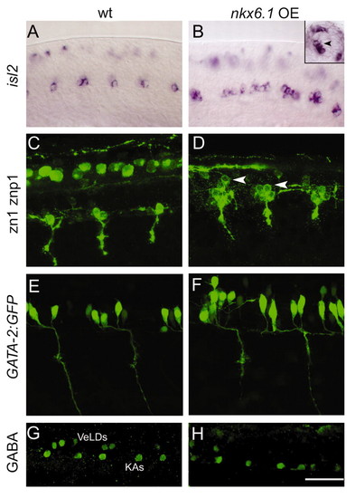
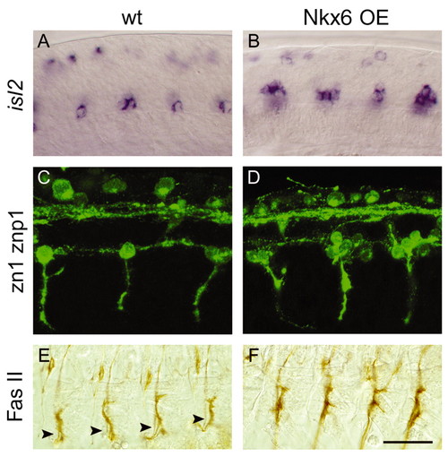
nkx6.1 is sufficient to induce zebrafish motoneurons and repress interneurons. (A) isl2-positive CaPs and VaPs at 18 hpf. Wild-type embryos have 1.3±0.7 isl2-positive cells per spinal hemisegment (n=5). (B) Embryos injected with nkx6.1 mRNA (Nkx6.1 OE) display supernumerary isl2-positive cells (2.3±0.5 per spinal hemisegment; n=7). These numbers are significantly different (P<0.001). Most RBs are not in focus in these images. Inset is a cross-section indicating the more dorsal position of supernumerary PMNs (arrowhead). (C) 24 hpf wild type labeled with zn1 and znp1 antibodies reveals 1 or 2 CaP/VaP cell bodies and axons as well as RBs. (D) Embryos injected with nkx6.1 RNA have 3-4 CaP/VaPs with normal axons, but some are more dorsally located (arrowheads). RBs are out of focus. (E) Wild-type 24 hpf GATA-2:GFP embryo has clusters of 3.0±1.7 SMNs in each rostral spinal cord hemisegment (8 hemisegments in 2 embryos). (F) nkx6.1 RNA-injected embryos have 4.0±2.8 SMNs per cluster (n=4; numbers are significantly different, P<0.009). All views are lateral, rostral to left. Scale bar: 50 μm for A,B; 33 μm for C-H. EXPRESSION / LABELING:
|
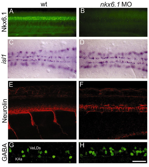
Nkx6.1 is required for zebrafish secondary motoneuron formation. (A) Nkx6.1 protein in 24 hpf wild-type spinal cord. (B) nkx6.1 MO-injected embryo has no detectable protein in spinal cord at 24 hpf. (C) Wild-type and (D) MO-injected 12 hpf embryos have the same pattern of isl1-positive PMNs. RBs are out of focus on each side of PMNs. (E) At 48 hpf, ventral spinal cord is full of Neurolin-positive SMNs. (F) MO-injected embryo has few remaining SMNs (75% of injected animals, n=565). The brightly-stained cuboidal cells are floorplate. (G) GABA antibody labels KA and VeLD interneurons in ventral spinal cord at 24 hpf. (H) In MO-injected embryos there is a dramatic increase in GABA-positive VeLD interneurons (n=21). Rostral to left in all images. A,B,E-H lateral views. C,D dorsal views Scale bar: 50 μm for A-D; 33 μm for E-H. EXPRESSION / LABELING:
PHENOTYPE:
|
Nkx6 function is conserved between fish and flies. (A) isl2-positive CaPs and VaPs in an18 hpf zebrafish embryo. isl2-positive RBs are out of focus. Wild-type embryos have 1.3±0.7 isl2-positive motoneurons per spinal hemisegment (n=5). (B) 18 hpf embryo ectopically expressing Nkx6 (fly gene, Nkx6 OE) RNA with supernumerary isl2-positive cells (2.3±0.3 isl2-positive cells per hemisegment), similar to the phenotype caused by ectopic expression of zebrafish nkx6.1 RNA (compare with Fig. 7B). The number of isl2-positive motoneurons is significantly different (P<0.0003). (C) PMNs in 24 hpf wild-type embryo labeled with zn1 and znp1; RBs and several axon tracts are also visible. (D) 24 hpf embryo ectopically expressing Nkx6 (fly gene) RNA generates ectopic PMNs (compare with Fig. 7D). (E) Stage 17 wild-type fly embryo stained with Fas II. Arrowheads indicate SNb nerves. (F) sca>Nkx6.1 (fish gene) fly embryos with visibly thicker SNb nerves (88%, n=42 hemisegments). (A-D) Lateral views of zebrafish spinal cord. (E,F) Dissected fly embryo musculature. Scale bar: 50 μm for A,B; 33 μm for C-F. EXPRESSION / LABELING:
|

Unillustrated author statements |