
Fig. 4
- ID
- ZDB-FIG-100506-2
- Publication
- Cheesman et al., 2004 - Zebrafish and fly Nkx6 proteins have similar CNS expression patterns and regulate motoneuron formation
- Other Figures
- All Figure Page
- Back to All Figure Page
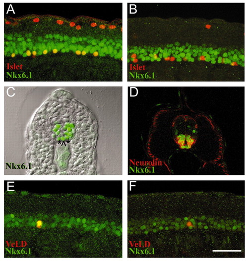
Nkx6.1 is expressed in zebrafish motoneurons. Nkx6.1 (green) Islet1/2 (red) antibody double labels. (A) 14 hpf lateral view reveals Rohon-Beard neurons (RBs) in dorsal spinal cord express only Islet proteins and PMNs in ventral spinal cord are double labeled (yellow). (B) By 18 hpf Nkx6.1 is largely absent from PMNs; most RBs are out of focal plane. (C) Cross-section of 24 hpf embryo showing Nkx6.1-positive nuclei, which comprise about five cell rows in the ventral spinal cord including medial (^) and lateral (*) floorplate. (D) Cross-section of 48 hpf embryo double labeled with Nkx6.1 (green) and Neurolin (red) antibodies. Nkx6.1-positive nuclei are surrounded by Neurolin-labeled cell surfaces. Laterally-located slow muscle cells are also Neurolin-positive. (E) 20 hpf lateral view of individually labeled VeLD interneuron (yellow) expressing Nkx6.1 (green). Most individually labeled VeLDs in 20-22 hpf embryos expressed Nkx6.1 (n=11, 82%). (F) By 24 hpf VeLDs have downregulated Nkx6.1 (n=13, 85%). Scale bar: 50 μm for A,B,D,E,F; 75 μm for C. |