
- Title
-
Intraspinal serotonergic neurons consist of two, temporally distinct populations in developing zebrafish
- Authors
- Montgomery, J.E., Wiggin, T.D., Rivera-Perez, L.M., Lillesaar, C., Masino, M.A.
- Source
- Full text @ Dev. Neurobiol.
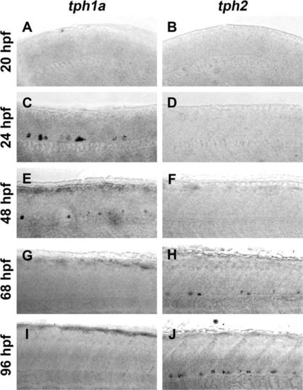
Expression of tph1a precedes expression of tph2 in the developing spinal cord. Spinal expression of tph1a (A, C, E, G, and I) and tph2 (B, D, F, H, and J) were compared by in situ hybridization in whole-mount embryos and larvae. A and B: Whole-mount in situ hybridization did not detect tph1a (A) or tph2 (B) transcripts in 20 hpf larvae. C-F: At 24 and 48 hpf tph1a (C and E), but not tph2 (D and F), was detected in the ventral spinal cord. G-J: Expression of tph1a was not observed after 68 hpf (G and I). Tph2-expressing cells were observed in the ventral spinal cord at 68 hpf (H) and labeling became more prominent at 96 hpf (J). |
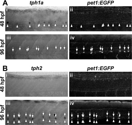
Spinal expression of pet1:EGFP corresponds to tph2, but not tph1a expression. Transcripts for tph1a (A) and tph2 (B) were detected by in situ hybridization (arrowheads) and EGFP was detected by immunohistochemistry (arrows) in whole-mount Tg(-3.2pet1:EGFP)ne0214 larvae. A: At 48 hpf, in situ hybridization revealed tph1a mRNA that was localized to cells distributed along the ventral spinal cord (Ai) and preceded pet1:EGFP transgene expression (Aii). Spinal tph1a expression was not observed at 96 hpf (Aiii) and EGFP was detected in cells in the ventral spinal cord (Aiv). B: Neither tph2 nor pet1:EGFP was expressed at 48 hpf (Bi and Bii). At 96 hpf, the majority of labeled cells were double-positive for tph2 and pet1:EGFP (Biii and Biv; Colabeling indicated by the presence of vertically aligned arrowheads and arrows). Black arrows indicate a single cell that expressed pet1:EGFP, but not tph2. |
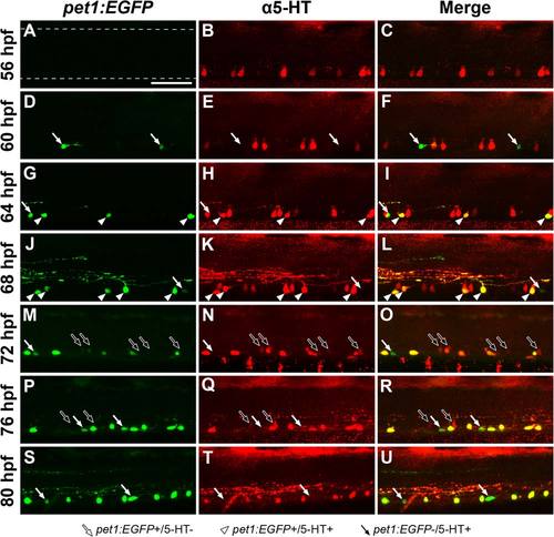
Spinal 5-HT antibody labeling transitions from pet1:EGFP- to pet1:EGFP+ cells during development. Tg(-3.2pet1:EGFP)ne0214 larvae were labeled with antibodies to 5-HT every 4 h between 56 and 80 hpf. Confocal Z-stacks were collected from the midbody region of the spinal cord (centered on body segment 15; dashed lines in A represent the dorsal and ventral boundaries of the spinal cord, approximately the same location in all panels). White arrows indicate cells that expressed pet1:EGFP and were not labeled with 5-HT antibodies, white arrowheads indicate cells in which pet1:EGFP expression and 5-HT colocalized, and black arrows indicate cells that did not express pet1:EGFP but were faintly labeled with 5-HT antibodies. A-C: 5-HT (B) was detected in cells in the ventral spinal cord before expression of pet1:EGFP (A, merge in C). D-F: Pet1:EGFP expression (D) was initially observed at 60 hpf in cells that did not colabel with 5-HT (E, merge in F; white arrows). G-L: At 64 and 68 hpf, a subset of pet1:EGFP+ cells (G, J) were 5-HT+ (H, K, merge in I, L; white arrowheads). 5-HT antibodies continued to detect pet1:EGFP- cells in the ventral spinal cord at 64 and 68 hpf (red cells in merged panels I, L). M-R: Colabeling of cells with pet1:EGFP (M, P) and 5-HT antibodies (N, Q) continued at 72 and 76 hpf (merge in O, R). The number of pet1:EGFP-/5-HT+ cells detected, and the intensity of 5-HT antibody labeling in these cells, were both reduced (black arrows). S-U: 5-HT antibody labeling (T) of cell somas was restricted to pet1:EGFP+ cells (S) in the ventral spinal cord at 80 hpf (merge in U). Scale bar = 50 µm. |
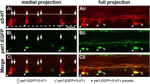
ISNs that express pet1:EGFP are morphologically distinct from those that do not express pet1:EGFP. A 64 hpf Tg(-3.2pet1:EGFP)ne0214 larva was labeled with antibodies to 5-HT, mounted laterally, and a confocal Z-stack was collected through the mediolateral extent of the spinal cord. The morphological structure of ISN somas (red) was examined in a confocal projection restricted to the medial spinal cord (Ai-Ci, medial projection) and a projection through the entire width of the spinal cord (Aii-Cii; full projection). A: 5-HT was detected in cell bodies near the ventral boundary of the spinal cord (dashed line, same position in all panels) that possessed two distinct shapes; dorsoventrally elongated conical cell bodies with apical terminals (Ai, arrows) and round or ovoid cell bodies (Aii, white arrowheads) that each possessed a single, prominent projection (Aii, black arrowheads). Apical terminals of the conically-shaped cells were positioned near the central canal (represented by a dotted line, same position in all panels). B and C: Pet1:EGFP (B, green) was expressed in ISNs with ovoid (white arrowheads), but not conical somas (arrows; merge in C). Scale bar = 20 µm. |
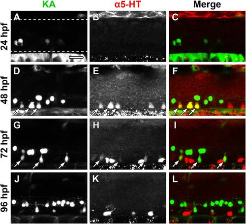
Figure 6. Kolmer-Agduhr (KA) neurons are 5-HT immunoreactive at 48 and 72 hpf. The Et(-1.5hsp70l:Gal4-VP16)s1003t and Tg(UAS-E1b:Kaede)s1999t lines were crossed to express Kaede in KA neurons (green) of progeny, which were immunolabeled for 5-HT (red). The Et(-1.5hsp70l:Gal4-VP16)s1003t line did not drive transgene expression in all KA neurons. A-C: Kaede expression (A), but not 5-HT antibody labeling (B, merge in C), was detected at 24 hpf (dashed lines represent the dorsal and ventral boundaries of the spinal cord, approximately the same location in all panels). D-F: A subset of ventral KA neurons (D) were labeled with 5-HT antibodies (E, merge in F; arrows indicate colocalization). G-I: 5-HT antibody labeling (H) of KA neurons (G) was faint at 72 hpf (arrows) and Kaede-negative cells were more strongly labeled with 5-HT antibodies (H, merge in I). J-L: Kaede expression (J) and 5-HT antibody labeling (K) did not overlap at 96 hpf (L). Scale bar = 20 µm. |
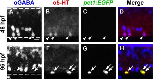
GABA and 5-HT immunolabeling overlap in ISNs that do not express pet1:EGFP. Whole-mount Tg(-3.2pet1:EGFP)ne0214 larvae were double-labeled with antibodies to GABA (blue) and 5-HT (red; dashed lines represent the dorsal and ventral spinal cord boundaries, same in A-D and E-H). A-D: A subset of ventral GABA-positive neurons (A) colabeled with 5-HT antibodies (B, merge in D; arrowheads indicate colocalization of GABA and 5-HT) at 48 hpf. Expression of pet1:EGFP (green) was not detected at 48 hpf (C). E-H: GABA (E) ceased to colocalize with 5-HT (F) at 96 hpf, as 5-HT became restricted to non-GABAergic pet1:EGFP+ (G; merge in H; arrows indicate colocalization of pet1:EGFP and 5-HT). Scale bar = 10 µm. |
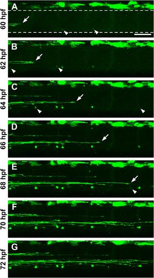
Visualization of developing spinal pet1:EGFP+ neurons and descending raphe projections. An anesthetized 60 hpf Tg(-3.2pet1:EGFP)ne0214 larva was embedded laterally in agarose. Images centered on the midbody (body segment 15) were collected using a confocal microscope every two hours between 60 and 72 hpf. The larva was maintained at 28.5°C when not being imaged. Arrows indicate the most caudal descending raphe projection at each age and arrowheads indicate pet1:EGFP+ cells as they first became visible. A: At 60 hpf, raphe projections were present in the most rostral region of the field of view (arrow). Two faintly pet1:EGFP+ cells were visible in the ventral spinal cord (A; dashed lines represent the dorsal and ventral boundaries of the spinal cord, approximately same position in all panels). B and C: Raphe projections extended further caudally (arrows) and additional cell bodies were detected (arrowheads) at 62 (B) and 64 hpf (C). D and E: Raphe projections continued to grow in a caudal direction at 64 and 68 hpf (D and E, arrows) and one additional cell was observed (arrowhead) at 68 hpf (E). F and G: Raphe fibers extended across the entire field of view and no new cells were detected at 70 (F) and 72 (G) hpf. Scale bar = 50 µm. |
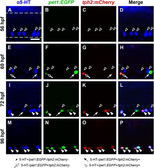
Spinal 5-HT immunoreactivity precedes both pet1:EGFP and tph2:mCherry transgene expression, and later becomes restricted to cells that coexpress pet1:EGFP and tph2:mCherry. Wholemount Tg(-3.2pet1:EGFP)ne0214 and Tg(tph2:nfsB-mCherry)y226 double transgenic larvae were labeled with antibodies to 5-HT (blue) and confocal images were collected from the midbody region of the spinal cord (dashed lines in A represent the dorsal and ventral boundaries of the spinal cord, approximately same position in all panels). Black arrowheads indicate cells that are 5-HT+ but did not express either transgene, white arrows indicate cells that were positive for only pet1:EGFP expression (green), black arrows indicate a cell that coexpressed pet1:EGFP and tph2:nfsB-mCherry (tph2:mCherry; red) but did not contain 5-HT, and white arrowheads indicate cells that were positive for all three markers. A-D: Antibodies to 5-HT revealed cells in the ventral 48 hpf spinal cord (A, black arrowheads) before pet1:EGFP (B) or tph2:mCherry (C) expression was detected (merge in D). E-H: At 60 hpf, 5-HT antibody labeling (E) that did not overlap with pet1:EGFP (F) or tph2:mCherry (G) expression persisted (black arrowheads). 5-HT- cells that expressed only pet1:EGFP (white arrows) or coexpressed pet1:EGFP and tph2:mCherry (black arrows) were observed in the ventral spinal cord (merge in H). I-L: At 72 hpf, pet1:EGFP+ cells (J) colabeled with tph2:mCherry expression (K) and 5-HT (I, white arrowheads), although a portion did not colabel (white arrow). 5-HT antibody labeling continued, but was reduced in cells that did not express either transgene (black arrowheads). M-P: By 96 hpf, 5-HT antibody labeling (M) was restricted to cells that coexpressed pet1:EGFP (N) and tph2:mCherry (O, merge in P, white arrowheads). Because pet1:EGFP was expressed in cells that did not coexpress tph2:mCherry (H and L, white arrows), but tph2:mCherry was only detected in pet1:EGFP+ cells (H, L, and P, black arrows and white arrowheads), expression from the pet1 promoter likely preceded expression from the tph2 promoter. Further, 5-HT was detected in cells that coexpressed both transgenes (L and P, white arrowheads) and not in cells that expressed only pet1:EGFP (H and L, white arrows). This suggested that 5-HT synthesis occurred after tph2:mCherry expression. Scale bar = 20 µm. |
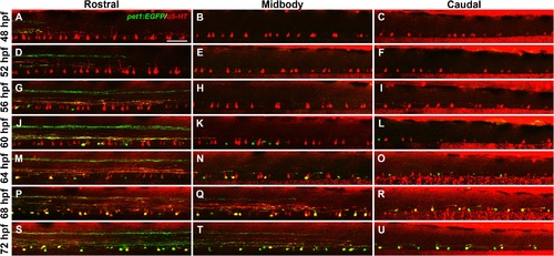
5-HT antibody labeling of ISNs progresses in a rostral to caudal direction. Tg(- 3.2pet1:EGFP)ne0214 larvae between the ages of 48 and 72 hpf were labeled with antibodies to 5-HT (red) and imaged in regions of the spinal cord centered on body segments 7 (rostral), 15 (midbody), and 23 (caudal). A-F: 5-HT was detected in ventral spinal cells in all regions at 48 (A-C) and 52 hpf (D-F). Expression of pet1:EGFP (green) was only observed in descending raphe processes in the rostral spinal cord (A and D). G-I: pet1:EGFP+ cells that did not colocalize with 5-HT were observed in the rostral region (G), but not the midbody (H) or caudal (I) region at 56 hpf. J-L: At 60 hpf pet1:EGFP+ cells were detected in the midbody (K) and caudal (L) regions and began to colocalize with 5-HT in the rostral region (J, yellow cells). M-O: A subset of pet1:EGFP+ cells were labeled with 5-HT antibodies in the rostral (M) and midbody (N) regions, but not in the caudal region (O), at 64 hpf. P-U: 5-HT antibody labeling continued to colocalize with pet1:EGFP expression (yellow), but was reduced in pet1:EGFP- cells (red cells) in all spinal cord regions at 68 (P-R) and 72 hpf (S-U). Projections from the raphe extended to the midbody region (Q and T) but had not reached the caudal spinal cord (R and U) at 68 and 72 hpf. Scale bar = 50 µm. |