
Fig. 2
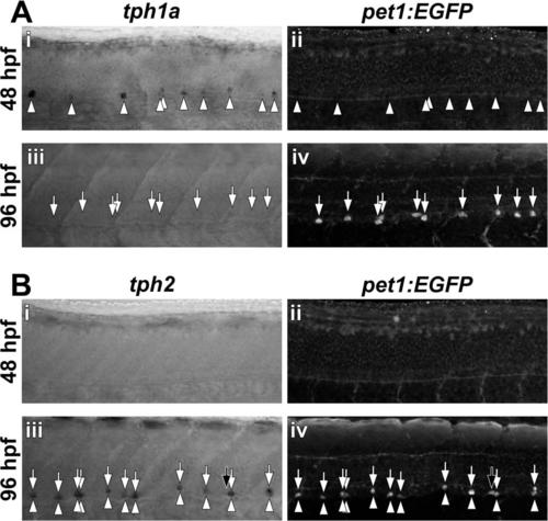
Spinal expression of pet1:EGFP corresponds to tph2, but not tph1a expression. Transcripts for tph1a (A) and tph2 (B) were detected by in situ hybridization (arrowheads) and EGFP was detected by immunohistochemistry (arrows) in whole-mount Tg(-3.2pet1:EGFP)ne0214 larvae. A: At 48 hpf, in situ hybridization revealed tph1a mRNA that was localized to cells distributed along the ventral spinal cord (Ai) and preceded pet1:EGFP transgene expression (Aii). Spinal tph1a expression was not observed at 96 hpf (Aiii) and EGFP was detected in cells in the ventral spinal cord (Aiv). B: Neither tph2 nor pet1:EGFP was expressed at 48 hpf (Bi and Bii). At 96 hpf, the majority of labeled cells were double-positive for tph2 and pet1:EGFP (Biii and Biv; Colabeling indicated by the presence of vertically aligned arrowheads and arrows). Black arrows indicate a single cell that expressed pet1:EGFP, but not tph2. |