
Fig. S3
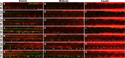
5-HT antibody labeling of ISNs progresses in a rostral to caudal direction. Tg(- 3.2pet1:EGFP)ne0214 larvae between the ages of 48 and 72 hpf were labeled with antibodies to 5-HT (red) and imaged in regions of the spinal cord centered on body segments 7 (rostral), 15 (midbody), and 23 (caudal). A-F: 5-HT was detected in ventral spinal cells in all regions at 48 (A-C) and 52 hpf (D-F). Expression of pet1:EGFP (green) was only observed in descending raphe processes in the rostral spinal cord (A and D). G-I: pet1:EGFP+ cells that did not colocalize with 5-HT were observed in the rostral region (G), but not the midbody (H) or caudal (I) region at 56 hpf. J-L: At 60 hpf pet1:EGFP+ cells were detected in the midbody (K) and caudal (L) regions and began to colocalize with 5-HT in the rostral region (J, yellow cells). M-O: A subset of pet1:EGFP+ cells were labeled with 5-HT antibodies in the rostral (M) and midbody (N) regions, but not in the caudal region (O), at 64 hpf. P-U: 5-HT antibody labeling continued to colocalize with pet1:EGFP expression (yellow), but was reduced in pet1:EGFP- cells (red cells) in all spinal cord regions at 68 (P-R) and 72 hpf (S-U). Projections from the raphe extended to the midbody region (Q and T) but had not reached the caudal spinal cord (R and U) at 68 and 72 hpf. Scale bar = 50 µm. |