
Fig. 1
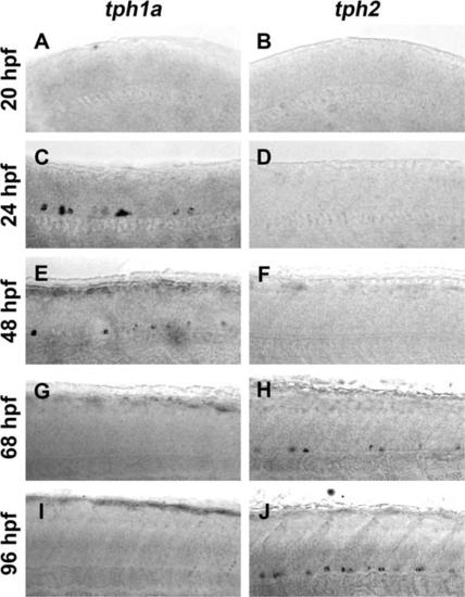
Expression of tph1a precedes expression of tph2 in the developing spinal cord. Spinal expression of tph1a (A, C, E, G, and I) and tph2 (B, D, F, H, and J) were compared by in situ hybridization in whole-mount embryos and larvae. A and B: Whole-mount in situ hybridization did not detect tph1a (A) or tph2 (B) transcripts in 20 hpf larvae. C-F: At 24 and 48 hpf tph1a (C and E), but not tph2 (D and F), was detected in the ventral spinal cord. G-J: Expression of tph1a was not observed after 68 hpf (G and I). Tph2-expressing cells were observed in the ventral spinal cord at 68 hpf (H) and labeling became more prominent at 96 hpf (J). |