
Fig. 6
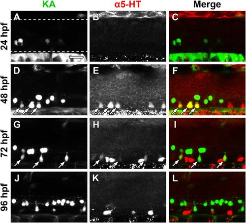
Figure 6. Kolmer-Agduhr (KA) neurons are 5-HT immunoreactive at 48 and 72 hpf. The Et(-1.5hsp70l:Gal4-VP16)s1003t and Tg(UAS-E1b:Kaede)s1999t lines were crossed to express Kaede in KA neurons (green) of progeny, which were immunolabeled for 5-HT (red). The Et(-1.5hsp70l:Gal4-VP16)s1003t line did not drive transgene expression in all KA neurons. A-C: Kaede expression (A), but not 5-HT antibody labeling (B, merge in C), was detected at 24 hpf (dashed lines represent the dorsal and ventral boundaries of the spinal cord, approximately the same location in all panels). D-F: A subset of ventral KA neurons (D) were labeled with 5-HT antibodies (E, merge in F; arrows indicate colocalization). G-I: 5-HT antibody labeling (H) of KA neurons (G) was faint at 72 hpf (arrows) and Kaede-negative cells were more strongly labeled with 5-HT antibodies (H, merge in I). J-L: Kaede expression (J) and 5-HT antibody labeling (K) did not overlap at 96 hpf (L). Scale bar = 20 µm. |