
Fig. 5
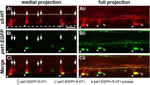
ISNs that express pet1:EGFP are morphologically distinct from those that do not express pet1:EGFP. A 64 hpf Tg(-3.2pet1:EGFP)ne0214 larva was labeled with antibodies to 5-HT, mounted laterally, and a confocal Z-stack was collected through the mediolateral extent of the spinal cord. The morphological structure of ISN somas (red) was examined in a confocal projection restricted to the medial spinal cord (Ai-Ci, medial projection) and a projection through the entire width of the spinal cord (Aii-Cii; full projection). A: 5-HT was detected in cell bodies near the ventral boundary of the spinal cord (dashed line, same position in all panels) that possessed two distinct shapes; dorsoventrally elongated conical cell bodies with apical terminals (Ai, arrows) and round or ovoid cell bodies (Aii, white arrowheads) that each possessed a single, prominent projection (Aii, black arrowheads). Apical terminals of the conically-shaped cells were positioned near the central canal (represented by a dotted line, same position in all panels). B and C: Pet1:EGFP (B, green) was expressed in ISNs with ovoid (white arrowheads), but not conical somas (arrows; merge in C). Scale bar = 20 µm. |