
|
Fig. 7
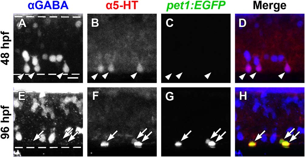
GABA and 5-HT immunolabeling overlap in ISNs that do not express pet1:EGFP. Whole-mount Tg(-3.2pet1:EGFP)ne0214 larvae were double-labeled with antibodies to GABA (blue) and 5-HT (red; dashed lines represent the dorsal and ventral spinal cord boundaries, same in A-D and E-H). A-D: A subset of ventral GABA-positive neurons (A) colabeled with 5-HT antibodies (B, merge in D; arrowheads indicate colocalization of GABA and 5-HT) at 48 hpf. Expression of pet1:EGFP (green) was not detected at 48 hpf (C). E-H: GABA (E) ceased to colocalize with 5-HT (F) at 96 hpf, as 5-HT became restricted to non-GABAergic pet1:EGFP+ (G; merge in H; arrows indicate colocalization of pet1:EGFP and 5-HT). Scale bar = 10 µm.