
- Title
-
Emilin genes are duplicated and dynamically expressed during zebrafish embryonic development
- Authors
- Milanetto, M., Tiso, N., Braghetta, P., Volpin, D., Argenton, F., and Bonaldo, P.
- Source
- Full text @ Dev. Dyn.
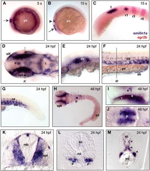
Whole-mount in situ hybridization of zebrafish embryos, visualizing the expression pattern of emilin1a at different developmental stages. A: Expression in the midbrain (arrow) at 5 somites (s). B: Expression in the midbrain (arrow) and in the first rhombomere (arrowhead) of hindbrain at 15 s. C: Double staining with egr2b probe (in red), labeling the third and fifth rhombomeres of hindbrain, confirms emilin1a expression in the dorsal side of first rhombomere and in the midbrain tectum at 15 s. D: Expression in epiphysis and head mesenchyme, especially in the territories of the olfactory and optic primordia, at 24 hours postfertilization (hpf). E: Expression in the otic vesicle at 24 hpf. F,G: Expression in the circulatory system of the trunk (F) and tail (G) at 24 hpf. H-J: Low (H) and high (I,J) magnifications of 48 hpf embryos, showing expression of emilin1a in epiphysis, sclera and retina (H,I), and in the pronephros (H,J). K: Cross-section of a 24 hpf embryo (level of section is marked by a dotted line in D), showing expression of emilin1a in the head mesenchyme. L: Cross-section of a 24 hpf embryo, showing expression in the pronephros. M: Cross-section of a 24 hpf embryo (level of section is marked by a dotted line in F), showing expression in the arterial, venous, and intersegmentary blood vessels (arrowheads). Except for embryos in panels A, B, and M, yolk was removed and embryos were mounted between glass coverslips. In A-J, anterior side of embryo is on the left; in the cross-sections, dorsal side is at the top. A-C and E-G are lateral views; D, H, and I are dorsal views; J is a ventral view. ab, angioblasts; ca, caudal aorta; cv, cardinal vein; da, dorsal aorta; e, epiphysis; i, isthmus; mb, midbrain; no, notochord; olf, olfactory placode; opt, optic cup; ov, otic vesicle; p, pronephros; pcv, posterior cardinal vein; r1-r5, first-fifth rhombomeres; r, retina; s, sclera; sc, spinal cord; t, tectum; yo, yolk. EXPRESSION / LABELING:
|
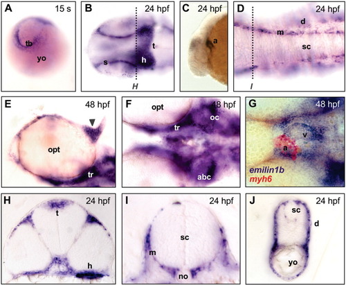
Whole-mount in situ hybridization for emilin1b in zebrafish embryos at different developmental stages. A: Expression in the tail bud at 15 somites (s). B: Expression in the sclera and in the midbrain at 24 hours postfertilization (hpf); a darker area, corresponding to labeling of the heart, is also visible. C: Lateral view, showing expression in the developing heart at 24 hpf. D: Expression in meninges at 24 hpf. E: Expression in the cartilages of epiphyseal bar (arrowhead) and trabeculae cranii at 48 hpf. F: Expression in the developing neurocranium at 48 hpf. G: Double hybridization with emilin1b (blue) and myh6 (red) probes, confirming expression in the heart at 48 hpf. H,I: Cross-sections of a 24 hpf embryo (levels of sections are marked by dotted lines in B and D), showing expression in heart, midbrain tectum and meninges. J: Cross-section of a 24 hpf embryo, showing expression in dermis. Except for A, C, and J, embryos were dissected away from the yolk and flat mounted. In A, posterior side of embryo is at the top; in B-G, anterior side is on the left; in the cross-sections, dorsal side is at the top. B and D are dorsal views, E-G are ventral views, C is a lateral view. a, atrium; abc, anterior basicranial commissure; d, dermis; ep, ethmoid plate; h, heart; m, meninges; no, notochord; oc, otic capsule; opt, optic cup; pch, parachordal cartilage; s, sclera; sc, spinal cord; t, tectum; tb, tail bud; tr, trabeculae cranii; v, ventricle; yo, yolk. EXPRESSION / LABELING:
|
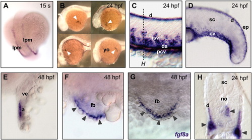
Whole-mount in situ hybridization for emilin2a in zebrafish embryos at different developmental stages. A: Expression in cells of the lateral mesoderm at 15 somites (s). B: Expression by cells in the yolk, corresponding to macrophage clusters (arrowheads), at 24 hours postfertilization (hpf). C,D: Expression in the circulatory system and in the dermis of trunk (C) and tail (D) at 24 hpf. E: Expression in the atrium of the developing heart at 24 hpf. F: Expression in the apical ectodermal ridge (arrowheads) of fin buds at 48 hpf. G: Staining of the apical ectodermal ridge (arrowheads) of fin buds with a fgf8a probe at 48 hpf, showing the same pattern of emilin2a. H: Cross-section of a 24 hpf embryo (level of section is marked by a dotted line in C), showing expression in the vasculature (arrowheads) and in the dermis. With the exception of A and B, the yolk was removed and embryos were flattened. A is a dorsal view, anterior side of embryo is at the top; B-G are lateral views, anterior side is on the left; in the cross-section, dorsal side is at the top. a, atrium; cv, caudal vein; d, dermis; da, dorsal aorta; ep, epidermis; fb, fin bud; isv, intersegmental blood vessels; lpm, lateral plate mesoderm; no, notochord; pcv, posterior cardinal vein; sc, spinal cord; ve, heart ventricle; yo, yolk. EXPRESSION / LABELING:
|
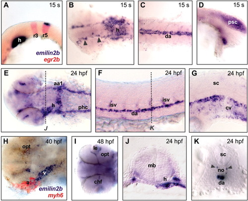
Whole-mount in situ hybridization for emilin2b in zebrafish embryos at different developmental stages. A: Expression in the heart primordium and in the dorsal side of fifth rhombomere of the hindbrain at 15 somites (s), as confirmed by double labeling with a egr2b antisense probe, that marks the third and fifth rhombomeres (in red). B: Expression in blood vessels of the head (arrowheads) and in the heart at 15 s. C: Expression in the dorsal aorta at 15 s. D: Expression in the posterior region of spinal cord at 15 s. E: Labeling of the heart and the circulatory system of the head at 24 hours postfertilization (hpf). F: Lateral view of trunk, showing expression in dorsal aorta and intersegmental blood vessels. G: Expression in the caudal vein at 24 hpf. H: Double hybridization with emilin2b (blue) and myh6 (red) probes, showing expression in the ventricle and part of the atrial chamber of heart at 40 hpf. I: At 48 hpf, emilin2b expression in the circulatory system becomes restricted to head vessels. J: Cross-section of a 24 hpf embryo (level of section is marked by a dotted line in E), showing expression in heart and circulatory system. K: Cross-section of a 24 hpf embryo (level of section is marked by a dotted line in F), showing expression in dorsal aorta and intersegmental blood vessels (arrowheads). All embryos were dissected away from the yolk and mounted between glass coverslips. In A-I, anterior side of embryo is on the left; in cross-sections, dorsal side is at the top. A,D,F and G are lateral views; B and C are dorsal views; E, H. and I are ventral views. aa1, mandibular arch; a, atrium; chf, choroid fissure; cv, caudal vein; da, dorsal aorta; h, heart; isv, intersegmental blood vessels; le, lens; mb, midbrain; no, notochord; opt, optic cup; phc, primordial hindbrain channel; psc, posterior spinal cord; r3, third rhombomere; r5, fifth rhombomere; sc, spinal cord; v, ventricle. EXPRESSION / LABELING:
|
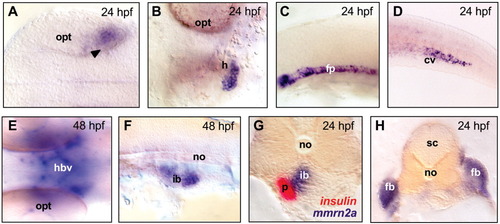
Whole-mount in situ hybridization for mmrn2a in zebrafish embryos at 24 hours postfertilization (hpf) and 48 hpf. A: Expression in the posterior retina (arrowheads) of the developing eye at 24 hpf. B: Ventral view, showing expression in the heart at 24 hpf. C,D: Expression in the anterior part of the floor plate (C) and in the caudal vein (D) at 24 hpf. E: Expression in the circulatory system of the head at 48 hpf. F: Expression in a territory corresponding to the developing intestinal bulb. G: Cross-section of a 24 hpf embryo after double labeling with insulin and mmrn2a probes; the insulin probe (in red) marks the β-cell precursors of pancreas, located behind the intestinal bulb, while mmrn2a labels the intestinal bulb but not the pancreas. H: Cross-section of a 24 hpf embryo, showing expression in the fin buds. All embryos were dissected away from the yolk and flattened. In A-F, anterior side of embryo is on the left; in the cross-sections, dorsal side is at the top. A and E are dorsal views, B is a ventral view, C,D,F are lateral views. cv, caudal vein; fb, fin bud; fp, floor plate; h, heart; hbv, head blood vessels; ib, intestinal bulb; no, notochord; opt, optic cup; p, pancreas; sc, spinal cord. |
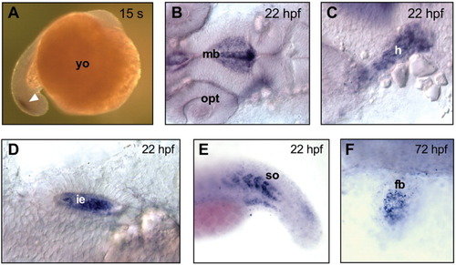
Whole-mount in situ hybridization for mmrn2b in zebrafish embryos at different developmental stages. A: Expression in the posterior region of the tail (arrow) at 15 somites (s). B: Expression at 22 hours postfertilization (hpf) in ependymal cells lining the midbrain ventricle. C: Expression in the heart at 22 hpf. D: Expression in the inner ear at 22 hpf. E: Expression in posterior somites at 22 hpf. F: Expression in the mesenchyme of fin buds at 72 hpf. Except for embryo in A, yolk was removed and embryos were mounted between glass coverslips. In A, anterior side of embryo is at the top; in B-F, anterior side is on the left. A and E are lateral views; B-D and F are dorsal view. fb, fin bud; h, heart; ie, inner ear; mb, midbrain; opt, optic cup; so, somites; yo, yolk. EXPRESSION / LABELING:
|
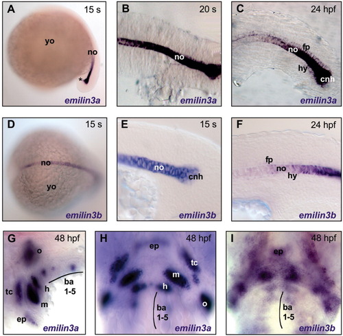
Whole-mount in situ hybridization for emilin3a and emilin3b in zebrafish embryos at different developmental stages. A-I: In situ hybridization was performed with probes for emilin3a (A-C,G,H) or emilin3b (D-F,I). A: Expression of emilin3a in the notochord and in the chordoneural hinge (asterisk) of the tail bud at 15 somites (s). B: strong labeling of the notochord by emilin3a at 20 s. C: At 24 hours postfertilization (hpf), expression of emilin3a decreases in the notochord and becomes evident in the floor plate of developing spinal cord and in the most posterior part of the hypochord. D: Labeling of the notochord by emilin3b at 15 s. E: Higher magnification of the posterior region of a 15 s embryo, showing emilin3b expression in the notochord and in the chordoneural hinge. F: At 24 hpf, expression of emilin3b in the notochord decreases and shows faint labeling of some cells in the floor plate and hypochord. G,H: Expression of emilin3a in the cartilage of branchial arches (G) and of craniofacial elements (H) at 48 hpf. I: Labeling of craniofacial mesenchyme by emilin3b at 48 hpf. Except for A and D, embryos were dissected away from the yolk and mounted between glass coverslips. A-C,E-G are lateral view, anterior side of embryo is on the left; D,H,I are dorsal view, anterior side is on the left in D and at the top in H and I. ba1-5, first-fifth branchial arches; cnh, chordoneural hinge; ep, ethmoid plate; fp, floor plate; h, hyoid cartilage; hy, hypochord; m, mandibular cartilage; no, notochord; o, opercular cartilage; tc, trabeculae cranii; yo, yolk EXPRESSION / LABELING:
|

Unillustrated author statements EXPRESSION / LABELING:
|