
|
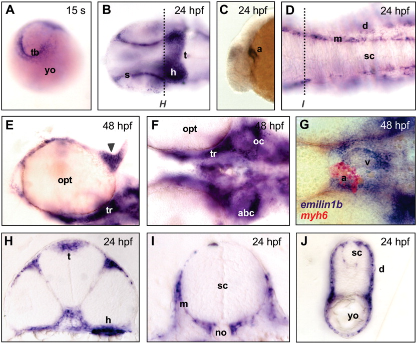
Fig. 3 Whole-mount in situ hybridization for emilin1b in zebrafish embryos at different developmental stages. A: Expression in the tail bud at 15 somites (s). B: Expression in the sclera and in the midbrain at 24 hours postfertilization (hpf); a darker area, corresponding to labeling of the heart, is also visible. C: Lateral view, showing expression in the developing heart at 24 hpf. D: Expression in meninges at 24 hpf. E: Expression in the cartilages of epiphyseal bar (arrowhead) and trabeculae cranii at 48 hpf. F: Expression in the developing neurocranium at 48 hpf. G: Double hybridization with emilin1b (blue) and myh6 (red) probes, confirming expression in the heart at 48 hpf. H,I: Cross-sections of a 24 hpf embryo (levels of sections are marked by dotted lines in B and D), showing expression in heart, midbrain tectum and meninges. J: Cross-section of a 24 hpf embryo, showing expression in dermis. Except for A, C, and J, embryos were dissected away from the yolk and flat mounted. In A, posterior side of embryo is at the top; in B-G, anterior side is on the left; in the cross-sections, dorsal side is at the top. B and D are dorsal views, E-G are ventral views, C is a lateral view. a, atrium; abc, anterior basicranial commissure; d, dermis; ep, ethmoid plate; h, heart; m, meninges; no, notochord; oc, otic capsule; opt, optic cup; pch, parachordal cartilage; s, sclera; sc, spinal cord; t, tectum; tb, tail bud; tr, trabeculae cranii; v, ventricle; yo, yolk.